Lower leg muscles
introduction

The lower leg is the part of the leg that is between the knee and the foot. The bony structures are made up of the tibia (Tibia) and fibula (Fibula), which in turn is formed by a tight band connection, the Membrana interossea cruris are connected. Below the knee, there is a tight joint between the tibia and fibula, a Amphiarthrosis, whereas the two lower leg bones are connected above the ankle joint and the so-called Syndesmosis tibiofibularis represent. The joint between the upper and lower leg (knee joint) is a hinge joint. It allows movements in the degrees of freedom, extension (Extension) and diffraction (Flexion), as well as rotational movements to a small extent. The shin-fibula connections (Art. Talofibularis proximales and distales) as plane joints only allow displacement movements, whereby the tibia-fibula joint distant from the body forms the ankle fork and thus stabilizes the upper ankle joint.
Classification of the lower leg muscles
The lower leg muscles are classified according to their function and location Muscles in two groups with two further subgroups each. The anterior lower leg muscles is divided into Extensors (Extensor) on the front side and in the Fibula muscleswhich is located in the area of the fibula on the outer side. The posterior lower leg muscles is divided into superficial flexors (Flexors), commonly known in anatomy as Triceps surae muscle are designated and in the deep flexors.
Front lower leg muscles
Extensors
The extensors of the anterior lower leg muscles are:
- Tibialis muscle anterior
- Extensor digitorum longus muscle and
- Extensor hallucis longus muscle.
The main function of the Tibialis anterior muscle is the Foot lift. Its tendon is deflected by ligaments of the ankle and ends on the inside of the foot in the middle of the arch, near the dorsum of the foot. With this approach the Tibialis anterior muscle the foot and especially the To lift the edge of the foot (Supination).
Of the Extensor digitorum longus muscle, also "long toe extension“Called, causes one Dorsiflexion (Pulling up) the second to fifth toes in the Metatarsophalangeal joint and of the foot in the ankle joint.
As "long big toe extensor“Is called the Extensor hallucis longus muscle, the one for that Pulling up the big toe responsible is. Depending on the position of the foot, it can also do that Inward or outward rotation of the lower ankle to support.
Fibula muscles
The fibula muscles include:
- Peroneus longus muscle
- Musculus fibularis brevis
As "long fibula muscle“Is called the Peroneus longus muscle. He sets similar to that Tibialis anterior muscle in the middle of the flexion of the foot, but there on the side of the sole of the foot. Its primary task is to Extend your foot towards the floor and to turn inwards. The tendon of the Peroneus longus muscle also gives the foot due to its transverse course Stability in the transverse arch.
Of the "short fibula muscles“Or Musculus fibularis brevis, cares like that Peroneus longus muscle for one Plantar flexion, so for stretching the foot down. In humans, its tendon runs in a common tendon sheath with that of des Peroneus longus muscle.
Lower leg muscles
Superficial layer
The superficial rear lower leg muscles include:
- Soleus muscle
- Gastrocnemius muscle
- Plantaris muscle
Work in the area of the lower leg muscles Soleus muscle and Gastrocnemius muscle close to each other. you are Synergists and are also used in anatomical terminology as Triceps surae muscle summarized.
Of the Soleus muscle (Clod muscle) is mostly from Gastrocnemius muscle covered, which is why it is only on the sides of the Lower leg is visible. His job is Plantar flexion, i.e. pulling the foot down towards the sole of the foot. He is also responsible for the Elevation of the inner edge of the foot while lowering the outer.
Of the Gastrocnemius muscle, also called the twin calf muscle, gives the human calf their characteristic shape. In close cooperation with the Soleus muscle it causes a in the upper ankle Pulling the foot down (Plantar flexion), one in the lower ankle Elevation of the inner edge of the foot (Supination) and in the knee joint a diffraction. Of the Gastrocnemius muscle possesses two muscle heads according to their location Caput mediale (inner head) and Caput laterale (outer head). Both arise at the lower part of the Thighbone. The Heel bone represents the approach of the two united muscle bellies. As Achilles tendon is called the common tendon of Gastrocnemius muscle and Soleus muscle.
A muscle that is small and regressed in humans, but still strongly developed in monkeys is the Plantaris muscle. Not present in every human being, this one shines muscle in the Plantar aponeurosis, a tendon plate in the area of the sole of the foot. The function of the Plantar muscle is almost in humans meaningless. He is only marginally involved in the flexion of the knee and the inward rotation of the lower leg in the flexed position.
Deep layer
The deep layer of the rear lower leg muscles includes:
- Tibialis posterior muscle
- Flexor hallucis longus muscle
- Flexor hallucis longus muscle
- Flexor digitorum longus muscle
Of the Tibialis posterior muscle, also known as the "posterior tibial muscle", attaches its tendon, which runs through the so-called tarsal tunnel, to the scaphoid and sphenoid bone. Its tasks are to lower (Plantar flexion) of the foot and the lifting of the inner edge of the foot.
Of the Flexor hallucis longus muscle, Latin for “long big toe flexor”, is the strongest of the deep toe flexor muscles. Its tendon crosses the tendon of the in the area of the sole of the foot Flexor digitorum longus muscle (see below). At this point there is a connection between the two flexor muscles, so that the Flexor hallucis longus muscle the effect of the Flexor digitorum longus muscle reinforced. In addition to the downward flexion of the big toe, the Flexor hallucis longus muscle the plantar flexion.
The "long toe flexor", Flexor digitorum longus muscle, bends all toes except the big toe towards the sole of the foot and supports the plantar flexion (flexion towards the sole of the foot) of the foot. Its tendon is divided into four tendons behind the tarsal tunnel, a canal in the area of the inner side of the ankle that is bounded by bone and connective tissue, which eventually reach the individual toes.
Fascia and boxes
As Fascia is called collagenous, fibrous connective tissuewhich joint- and Organ capsules educates and also Muscles, bone, Nerve tracts and encloses blood vessels. The entire lower leg muscles are called by the Fascia cruris surround. Individual muscle groups, depending on their function, are divided into different fasciae by further fasciae Compartments divided and separated from each other. This separation results in functional units in the anatomy Muscle boxes to be named.
The following are found in the lower leg Boxes:
- Extensor box: Tibialis anterior muscle, Extensor digitorum longus muscle, Extensor hallucis longus muscle
- Flexor box: Triceps surae muscle, Tibialis posterior muscle, Flexor hallucis longus muscle, Flexor digitorum longus muscle, Popliteus muscle
- Fibular islet: Musculus fibularis longus, Musculus fibularis brevis
The fascia, which Muscle tissue surround, cause a compressionthat can prevent swelling after use or injury.
Compartment syndrome

However, the anatomical separation of the individual muscle groups by fasciae carries the risk of bleeding into the muscle boxes after injuries. The compartment syndrome usually arises from trauma such as broken bones or as a result of blunt violence. Operations or muscular overload, as can occur in competitive and amateur athletes, can trigger a compartment syndrome through bleeding or edema formation.
Due to the reduced elasticity of the firm connective tissue of the fascia, the pressure within a muscle compartment can rise sharply, which also compresses the vascular nerve bundles of the lower leg. This is followed by an impairment of the blood supply and neuromuscular function. The early symptom is severe pain in the affected extremity combined with a feeling of tension and increasing sensory disorders such as numbness and tingling. The mobility can sometimes be severely restricted. Due to the increasing pressure in the box, the venous outflow is increasingly obstructed. If the arterial blood flow is initially preserved, a vicious circle begins, which leads to a further increase in pressure. As a result, the arterial blood flow comes to a standstill with an increasing lack of supply to the muscles. At this stage, motor deficits and a lack of pulse appear in the area after the affected muscle box. Due to the threat of extensive tissue destruction, the compartment syndrome is an absolute emergency that needs urgent care.
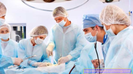
The treatment of choice for traumatic compartment syndrome is the surgical splitting of the fascia of the affected muscle compartment and the adjacent compartments in order to relieve pressure. After the swelling has subsided, the artificially made incision can either be sewn or covered with a skin graft. If the compartment syndrome is not treated in a timely manner, the affected muscle tissue can suffer massive destruction, which in extreme cases makes it necessary to amputate the extremity.
Read more on this topic at: Compartment syndrome
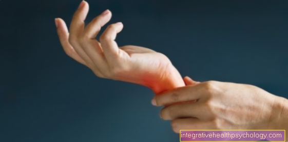
Shin splints
The Shin splints, too, depending on the localization medial (middle) resp. lateral (lateral) Tibial Edge Syndrome called, is a disease that usually occurs in connection with physical activity. It denotes load-dependent, dull or shooting pain on the edge of the shin. Are particularly at risk jogger or athletes who do intensive training in sports with particular stress on the shin or foot muscles.
Several conditions can trigger the pain. The exact Emergence of shin splints is not conclusive clarified. Pronation movements of the foot in particular, that is, lifting of the lateral edge of the foot with simultaneous lowering of the middle edge of the foot, seem to favor a tibial splint syndrome. According to the current state of knowledge, the causes are mostly Overuse of the muscles, Inflammation or injury to the muscle. Overuse can result from a variety of sports. However, the focus is on running or ball sports with a rapid change of direction such as football or handball. With untrained people Excessive exercise can quickly lead to shin splints. Even with experienced and trained athletes, it can result in a rapid weight gain or one Change of footwear get sick.
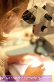
Often the pain occurs the day after exercise on. If you continue to train, the training can also be painful. At the point of heavy stress on the shin bone it can increase over time Periosteum inflammation for example the Shin come. Especially Amateur athlete tend to blame pain on muscle overuse. If an inflammation of the periosteum is suspected, an appropriate diagnosis using imaging methods (Magnetic resonance imaging) and subsequent therapy can be initiated. Athletes in particular find the sometimes long-term impairment in training, which is explained by the slow healing of the disease, particularly stressful.
The therapy itself is not infrequently unsatisfactory and the possibilities of medicine are sometimes limited. The focus is on Protection of the affected extremitywhich usually leads to a rapid improvement of the symptoms, which can, however, recur with renewed exercise. By local cooling the course of the disease can be influenced favorably. In severe pain you can Non-steroidal anti-inflammatory drugs how Ibuprofen® have a pain relieving effect. The doctor can provide assistance and advice when choosing insoles or supports that are worn in the shoe during training.
Congenital clubfoot
The innate clubfoot, also Pes equinovarus, is a Misalignment of the child's foot and occurs with a frequency of 1: 1000 births. J
ungen are affected twice as often as girls. Causal for foot deformity is one Disturbance of the balance of the lower leg muscleswhere the Plantar flexors, i.e. flexors of the foot towards the sole of the foot, and supinators, lifters of the middle edge of the foot, predominate.
As "Clubfoot muscle“Will also be Tibialis muscle posterior denotes the foot in Supination brings and bends towards the sole of the foot. The Misalignment is direct at birth and is a Combination of several deformities. As a rule, there is an inward rotation of the foot, the sickle foot position of the forefoot, equinus, Arches foot and a lateral deviation of the heel together.
The exact origin of the congenital clubfoot has not yet been clarified.
However, it is believed that the location of the embryo in the uterus is a crucial factor.
Also one decreased Amount of Amniotic fluid could encourage the development of clubfoot. As a result of a neural tube defect, the faulty formation of the embryonic system of the central Nervous system, paralysis of the lower leg muscles can lead to clubfoot. It is also discussed whether a congenital clubfoot is a result of ingestion Folic acid antagonists how Aminopterin® or Methotrexate® can occur in the fourth to twelfth week of pregnancy.
The treatment should be initiated immediately after birth. First there is the therapy in the Fixation of the foot, the so-called retention, which is gradually adapted to a correct position (redressement). The cast must be changed regularly, with the redressement continuing. At the age of about three months can a Operation on the Achilles tendon become necessary, whereby this is lengthened and the angle between the ankle and heel bone is corrected.
Further measures can be taken in the Transplant of Tibialis anterior muscle, Bone corrections or Joint stiffeners consist. To be shortened Muscles to stretch and the Joints to mobilize the foot physiotherapy procedures be used at an early stage, as it can lead to a renewed deviation into the deformity in the long term.