Tension pneumothorax
What is tension pneumothorax?
A tension pneumothorax is a special form of pneumothorax and represents a life-threatening injury to the lungs. In contrast to a collapsed lung (pneumothorax), a tension pneumothorax also has a type of valve mechanism in which more and more air accumulates in the chest, but which is not exhaled can be. There is an increasing increase in pressure within the chest and a shift in the mediastinum (the center of the chest, in which the heart and its supplying and draining blood vessels are located), endangering the cardiovascular system. The most common causes of tension pneumothorax are traumatic influences, such as traffic accidents.
Read more on the topic: Pneumothorax

causes
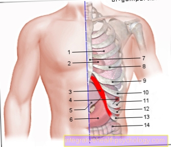
The causes of tension pneumothorax are the same as those of pneumothorax. The reason for this is that any pneumothorax can, through unfortunate circumstances, develop into a tension pneumothorax. The lungs are each enclosed by a thin skin, the pleura (pleura visceralis). Together with the pleura (pleura parietalis), which surrounds the ribs, the two form the so-called “pleural space”, in which there is a small amount of fluid and there is negative pressure. You can imagine it like two panes of glass that are damp and stick to each other.
If, for certain reasons, air enters the gap, the negative pressure disappears and the respective lung, which is only kept open by the negative pressure, collapses. This event is called a pneumothorax. In a tension pneumothorax, air enters the pleural space every time you inhale. When exhaling, however, this cannot escape again through a type of valve mechanism, whereupon the air becomes enriched.
The reason is a relocation of the entry point, for example through lung tissue. The causes of a pneumothorax and thus also of a tension tire are primarily traumatic events such as stab or gunshot injuries, broken ribs, bruises and overpressure in the lungs, for example from positive pressure ventilation or while diving. A spontaneous pneumothorax, for example when lifting heavy objects, is also possible. In addition, some medical interventions can injure the pleural space and thus lead to a doctor-related pneumothorax.
Read more about: Structure of the lungs
diagnosis
A tension pneumothorax is a very rapidly progressing event in which patients can worsen within a very short time. A clinical diagnosis is therefore often possible or necessary. This means that the rescue service or a doctor can suspect a tension pneumothorax based on external parameters such as pulse, blood pressure, skin color, breathing behavior and blocked blood vessels. A diagnosis is usually confirmed by a chest x-ray (chest x-ray) or, in rare cases, by a CT (computer tomography).
What can you see on the x-ray?
The chest X-ray, which usually takes a picture of the chest from the front as well as from the side, is the standard procedure for diagnosing tension pneumothorax. A pneumothorax is represented by one or, if necessary, two collapsed (collapsed) lungs and free air, which appears very dark. In the case of a tension tire, there is also a displacement of the mediastinum. This means that the heart and its supplying and draining blood vessels are shifted towards the healthy side. The more advanced the shift, the more dangerous the condition and the more urgent intervention is needed.
Concomitant symptoms
Accompanying symptoms of a tension pneumothorax are above all an increasing shortness of breath (Dyspnea), an increased heart rate (Tachycardia) and falling blood pressure (Hypotension), usually associated with symptoms of shock. The patients are also often very pale, suffer from dizziness, weakness, possible loss of consciousness and signs of cyanosis. Cyanosis describes a blue discoloration of the mucous membranes as a sign of an insufficient supply of oxygen.
Another symptom is a congestion of the blood vessels in the neck, which already indicates an advanced stage and a mediastinal displacement. Depending on the cause of the tension pneumothorax, there may also be pain in the chest and accompanying injuries.
Crackling under the skin
In medicine, crepitatio or crepitation is a crackling or crunching noise that can occur in different situations. With tension pneumothorax, crepitation can occur as part of a so-called skin emphysema. Skin emphysema is a superficial accumulation of air in the skin tissue. In the case of tension pneumothorax, this can result from external injury to the lungs. The air from the lungs escapes into the skin tissue and remains there.
Symptoms are the typical crepitatio, especially due to pressure on the corresponding areas, protrusions and the mobility of the respective protrusions. Skin emphysema is diagnosed by a chest X-ray.
Mediastinal displacement
Mediastinal displacement describes a displacement of the mediastinum towards the side of the healthy lung. The mediastinum is the center of the thorax, in which the heart and its supplying and draining blood vessels are located. The increasing pressure in the pleural space leads to a compression of the vessels supplying the heart (veins), which means that the heart is no longer supplied with sufficient blood and the blood pressure drops. This also leads to a congestion of the neck veins, which is an important symptom in diagnosis. In the course of the process, the increasing pressure also leads to compression of the heart and consequent cardiac arrest.
How is tension pneumothorax treated?
There are several different ways to relieve tension pneumothorax. It is important to release the pressure that builds up through the trapped air and thus counteract the mediastinal displacement. This is made possible either by a so-called relief puncture or by a chest drain. A relief puncture counteracts a tension pneumothorax and prevents the life-threatening condition.
In contrast to a chest drain, in which a tube is inserted into the pleural space and the original negative pressure is restored, a relief puncture is only an emergency measure and not a definitive therapy. Accordingly, relief is only used under enormous time pressure or in the rescue service. A needle is used to establish a connection between the pleural space and the outside air. The needle is inserted either on the upper edge of the second or third rib on the front of the chest (according to Monaldi) or on the upper edge of the fifth or sixth rib on the side of the chest (according to Bülau). It is important that you do not prick the lower edge of the rib, as this is where nerves and blood vessels run.
Read more on the topic: Chest drain
Duration and forecast
The duration and prognosis of a tension pneumothorax depends on when a valve mechanism has formed, how pronounced it is and how soon therapy has or can be carried out. If the valve mechanism is fully developed, a tension pneu develops within a few minutes, as approximately 500 ml of air enter the pleural space with each breath. Without therapy, tension pneumothorax is usually fatal as it leads to cardiac arrest.
If a tension tire is treated in good time and without complications, it will heal completely after a few days. However, this depends on concomitant injuries and the patient's state of health.
Can a tension pneumothorax be fatal?
A tension pneumothorax is an absolutely life-threatening condition and must be treated as early as possible. If there is no timely treatment, a tension pneumothorax is usually fatal. The reason is the compression of the mediastinum and the subsequent cardiac arrest. Unfortunately, a tension pneumothorax usually progresses very quickly, which makes urgent therapy all the more necessary. With timely and complication-free treatment without major accompanying injuries, however, a tension tire has a very good prognosis and usually heals very quickly.


