How can you measure intracranial pressure?
introduction
Under intracranial pressure - actually intracranial pressure (ICP) - one understands the pressure inside the skull, which is largely determined by the pressure in the liquor system. This consists of several cavities or Ventriclesin which the liquor, also known as "nerve water", washes the brain and spinal cord. A certain amount of pressure builds up. Since the intracranial pressure counteracts the pressure of the intracranial bleeding, it must not be too high. There are various methods of measuring intracranial pressure, which are presented below.
Here's what exactly increased intracranial pressure is

What is the normal intracranial pressure?
The intracranial pressure is usually measured in units of mmHg (millimeters of mercury) or cmH2O (centimeters of water). Values between 0 and 10 mmHg are considered normal, in some cases values up to 15 mmHg are stated as physiological. Values above 20 mmHg are in any case considered to be elevated. The higher the pressure, the more severe consequential damage can be.
What are the consequences of increased intracranial pressure?
Everyone knows that e.g. the finger swells when one is injured there. The problem with the brain, however, is that it sits in a rigid, bony shell. This protects it from injury, but also prevents it from expanding. If there is swelling (cerebral edema) due to an injury to the brain tissue, the brain can only expand minimally, the intracranial pressure rises relatively quickly and there is permanent pressure on the sensitive brain tissue. Other so-called space-consuming processes such as brain tumors, bleeding or brain abscesses can also lead to an increase in intracranial pressure. A short-term increase usually remains without long-term consequential damage, but typical symptoms are:
-
a headache
-
nausea
-
Impaired consciousness
-
unequal sized pupils (anisocoria)
-
Congestive papilla
-
many more possible!
A prolonged increase in intracranial pressure can lead to serious, permanent damage to the brain and must be avoided at all costs!
(ATTENTION: The intracranial pressure signs, especially headaches and nausea, are very unspecific and do not necessarily have to indicate increased intracranial pressure, they can also be symptoms of many other diseases! If you are unsure and these symptoms persist for a longer period of time for no apparent cause, you should however, to be on the safe side, consult your family doctor or neurologist!)
Feel free to read the main article on the topic as well Intracranial pressure sign
Severe consequences of acutely increased intracranial pressure are so-called herniations, i.e. entrapment of brain tissue. Depending on the location of the entrapment, a distinction is made primarily between:
-
an upper entrapment (entrapment of cerebellar parts)
-
a lower entrapment (entrapment of the brain stem)
The entrapment of the brain stem in particular is an often fatal consequence of increased intracranial pressure and must be treated immediately by emergency and intensive care!
How does intracranial pressure measurement work?
What methods are there?
In many cases, indications for measuring intracranial pressure are acute events such as unconscious unconsciousness, cerebral haemorrhage or severe infections such as meningitis or brain abscesses. But longer events such as a brain tumor or skull malformations are also possible causes of increased intracranial pressure. The intracranial pressure is measured in comatose patients and when there are signs of intracranial pressure: These include, above all, impaired consciousness, headaches, nausea, rounded pupils or abnormal breathing.
The intracranial pressure or intracranial pressure is the pressure that prevails in the cranial cavity and is composed of the blood pressure in the head and above all the CSF pressure.
The direct measurement is made invasively using a special probe with a diameter of 1-2 mm. To do this, the neurosurgeon first drills a hole in the bony skull and inserts the probe over it. This comes to rest in one of the following places:
-
over the meninges (epidural)
-
under the meninges (subdural)
-
in brain tissue (parenchymal)
-
in the liquor spaces (intraventricular)
This probe can now remain in place for several days. Since it is a very invasive procedure with many risks and possible complications, the patient should be placed in a specialized neurological monitoring or ideally intensive care unit.
Another possibility is to measure the CSF pressure as part of a lumbar puncture. The typical indication for this is idiopathic intracranial hypertension (obsolete: pseudotumor cerebri). With this disease, the CSF pressure has to be measured again and again and usually also reduced. For a lumbar puncture, a riser tube is connected to the puncture needle, with which the intracranial pressure can be approximately determined. Since the puncture needle has to be withdrawn after the puncture, no longer-term monitoring can of course be carried out.
In the fundus of the eye (Fundoscopy) the intracranial pressure cannot be measured, but a congestive papilla can be quickly and easily recognized as a sign of increased intracranial pressure. In the case of the papillae, the increased pressure in the skull - i.e. behind the eye - ultimately leads to a bulging of the optic nerve head in the eye.
Which doctor measures intracranial pressure?
The invasive measurement of intracranial pressure via a probe must be carried out by a neurologist or neurosurgeon in the hospital, ideally in a neurological intensive care or monitoring ward.
The measurement via the CSF puncture is also carried out by the neurologist, in children it can also be carried out by the pediatrician.
What is the brain pressure probe?
The 1-2 mm wide intracranial pressure probe is a special measuring device for measuring intracranial pressure. The probe is inserted during a neurosurgical operation. To do this, the surgeon first drills a hole in the skull and uses it to insert the probe. This is then either
-
over the meninges (epidural)
-
under the meninges (subdural)
-
in brain tissue (parenchymal)
-
or in the liquor spaces (intraventricular)
to lie down.
The probe itself is a liquid or air-filled catheter that converts the pressure to digitally as a pressure curve.
This is a very invasive procedure that can result in infections or injuries to the brain tissue, among other things. It is therefore essential to place the patient on a specialized neurological ward!
Here you can learn more about the anatomy of the Meninges and Liquor spaces
What does the ophthalmologist measure?
The ophthalmologist cannot measure the intracranial pressure, but he can prove an important intracranial pressure sign in the funduscopy: the congestive papilla. The increased pressure in the skull - that is, behind the eye - ultimately leads to the optic nerve head bulging in the eye. Usually this bulge is visible in both eyes.
If your ophthalmologist suspects you have increased intracranial pressure, you should urgently consult a neurologist for further clarification!
How can you measure intracranial pressure in a baby?
In the case of children and babies, it is generally important to avoid invasive examinations if possible. One advantage with babies is that the parts of the skull bone have not yet grown together and the fontanel is therefore open. Since there is no bone here, it is possible to detect signs of intracranial pressure in the skull with harmless ultrasound and thus indirectly detect increased intracranial pressure.
Can you also measure intracranial pressure with an MRI?
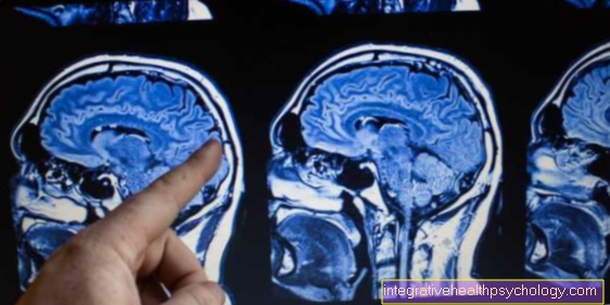
In the MRI, as in any imaging procedure of the skull, signs of intracranial pressure can be detected, and this includes above all
- pent-up, wide liquor spaces
- a displaced center line
- Transfer of liquor into the brain tissue (liquor diapedesis)
However, since there is no mechanical access to the inside of the skull, the exact pressure measurement cannot be determined in any imaging method.
Are there alternatives to measuring intracranial pressure?
As described above, intracranial pressure measurement is possible in various ways. If there is a reason, the measurement and monitoring should also always be carried out, as otherwise severe brain damage and death can result. The respective neurologist or neurosurgeon decides on the exact type of measurement on a case-by-case basis.


