How can I recognize breast cancer?
introduction
Especially at the beginning of breast cancer, when the tumor is still very small and in the early stages, there are often no noticeable signs. The tumor is often discovered by chance during a woman's self-scanning or routine examinations at the gynecologist.

The nodular change that can be felt is usually hard and fuzzy. Often it can no longer be shifted too well in the tissue because malignant tissue tumors have a tendency to grow together with the environment.
If the tumor is located behind the nipple, retraction of the nipple and the surrounding skin may also be noticeable. Furthermore, in some cases a change in the skin in the area above the tumor in the form of an orange peel-like texture can also be noticed. In advanced stages it can even lead to real ulceration (Ulceration) come.
In so-called inflammatory breast cancer - a special form of breast cancer - all signs of inflammation can be present (Redness, swelling, overheating, pain) and therefore often difficult to contract from a breast infection (mastitis) can be distinguished.
Please also read our article on this Swelling of the chest
Methods for examining the female breast are described below. The order is based on "invasiveness" (in medicine, methods are called invasive that penetrate the body) and the complexity of the investigation. The self-examination that is at the beginning is in no way stressful for the body and easy to carry out.
Self examination
The easiest method that female breast to examine it is to be palpated. 80% of breast cancer cases are still discovered by the affected women themselves.
Part of the statutory early detection program for Breast cancer is a Palpation of the chest from the age of 30. However, given the simplicity of this "method", its low cost and the complete absence of side effects, younger women should also take advantage of this option and examine themselves.
The rule is that not every hardening or change in the breast that can be felt also always means breast cancer. In 80% of the cases, the palpable nodular structures are benign changes.
method
About once a month a woman should look at her breasts in the mirror and feel them calmly. Stand in front of the mirror with your arms drooping. Look at her breasts from the front and the side. Pay attention to one-sided changes e.g. the surface of the skin, bulges, wrinkles, or retraction of the nipples.
Then the arms can be placed behind the head on both sides. The breasts should follow the movement and move upwards with it. Here, too, attention should be paid to retractions or unilateral changes in the shape of the breasts. New changes in the surface of the skin, protrusions or indentations are always a reason to arrange a check-up appointment with the gynecologist.
The best time to palpate the chest is about a week after the last menstrual period started. At this point the breast is particularly soft, later in the cycle the breast tissue becomes harder and knotty under the influence of the hormones.
After the onset of menopause, the breast can be palpated equally well at any point in time. If this is your first time examining your breast, don't be alarmed! Breast tissue consists not only of fat, but also of the mammary glands, palpable bumps and smaller bumps are normal. Changes over time are particularly important. So above all, pay attention to whether you are feeling any knots that weren't there a month ago.
When scanning the breasts, it is usually a matter of simple manipulations that allow a very precise overview of the nature of the breast tissue. The best way to palpate the chest is to take the arm on the same side behind your head. Another option is to place your hand under your chest and raise it slightly to create resistance when you feel.
The breast is divided into four quadrants by imagining a cross, the center of which is the nipple. For example, start on the upper inner quadrant and work your way with light circular movements from the outside to the inside, from quadrant to quadrant.
The fingertips of the index, middle and ring fingers are used to feel from the outside towards the nipple, always in the form of slightly powerful, small, circular movements. After that, you should examine the center of the breast around the nipple. Change the pressure your fingers exert, feel the surface and the depth of the tissue.
Feel your armpit and feel along the edge of the pectoral muscle and watch for changes, such as Knots or lumps, e.g. Do not let it move with light pressure.
In the end, you should take your nipple between your thumb and forefinger and pinch it gently. If you feel severe pain or observe the leakage of fluid or blood, you should make an appointment with your doctor.
You should also feel the area of the breast directly under the nipple and under the areola with light pressure. In order to be able to recognize changes more easily, this process should be repeated while lying down, just so the lower quadrants can be examined more easily.
Summary of the self-examination
- Viewing the chest with arms drooping
- Looking at the chest with both Poor behind the head
- Slowly scan all four quadrants with the arm behind the head or hand under the chest
- Palpation of the armpit and the edges of the Pectoral muscle
- Squeezing the nipple and touching the deeper tissue
- Repetition while lying down
Please also read our page Breast cancer screening
Info: breast cancer examination
The most common, namely about 55%, is found Breast cancer in the upper outer quadrant, so near the armpit. Cancer is found in about 15% of cases in the upper inner quadrant and in the area of the nipple. The lower inner quadrant is affected in 10% of all cases. The rarest of all, at 5%, is breast cancer in the lower outer quadrant.
Clinical signs of breast disease
Signs that can occur in breast cancer are described again in detail below. Give all the changes mentioned Indication of a breast disease. What kind of disease is your doctor needs to further clarify diagnostic means determine. If you notice any of the following changes, make an appointment with your gynecologist.
Please also read our page Signs of breast cancer
Breast lump
The most important clinical sign is the palpable, firm lump. With every palpable lump in the breast it must always be examined whether it is a malignant tumor or whether the diagnosis “breast cancer” can be ruled out.
The size of a lump can vary from the size of a pea to the size of a lime, depending on the stage of the cancer. Sometimes the lumps can be tender or cause a painful pulling sensation, but there are also findings that are completely painless.
Breast cancer nodules are usually fused with their surroundings, which is due to the decomposing growth of the cancer. They are therefore often difficult to move within the tissue and do not follow the pressure of the hand when scanning.
Depending on the size of the breasts and the lump, there may be a noticeable increase in the size of a breast. Irregularities in the size between the two breasts, which have always existed, are quite natural and do not require any further clarification. On average, the size of the lumps that are felt in the self-examination is a little over 2 cm. Mammography can detect nodes from a size of about 1 cm. However, due to the nature of the tissue, 15% of palpable tumors cannot be identified in the mammography, and therefore breast cancer cannot be identified.
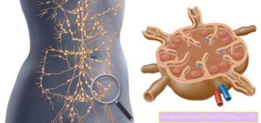
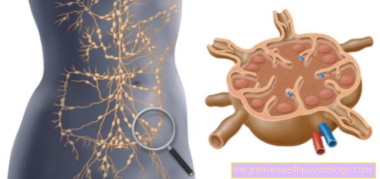
Lumps can also be felt on the edge of the chest muscle or in the armpits. These are then probably enlarged lymph nodes in the armpit. They are usually about the size of a lentil and usually cannot be felt. A distinction is made between benign and malignant enlarged lymph nodes. Benign enlargements occur in infectious diseases such as a simple cold but also with skin infections or various viral diseases. The enlargement is then due to the activation of the immune system. These lymph node swellings usually appear suddenly and the palpable lymph nodes feel soft, can be moved easily and are free from pressure. Malignant enlargements can e.g. occur in leukemia but also in other cancers (e.g. breast cancer). The lymph nodes can become very large and usually feel hard, are difficult to move against the environment and are sensitive to pressure.
Read more on the topic:
- Lymph node involvement in breast cancer
- Breast lump
Chest indentations and bulges
Lumps in the breast can lead to visible bulges due to their volume alone. More often, however, they lead to recoveries skin (also called plateau phenomena), which are usually particularly noticeable when the arm is raised. Retractions are caused by the tumor-related adhesions of Connective, Fat and skin tissue. Even very small knots, which are barely or not at all palpable, can lead to such adhesions and thus to indentations or bulges.
Orange peel on the chest
Orange skin, too Orange peel phenomenon or French peau d'oranges is a symptom that is more likely to occur in more advanced stages. The term vividly describes the change in the skin over the tumor. The Skin is slightly reddened and the pores are enlarged and emphasized. The orange skin is created by the accumulation of liquid in the skin, which causes it to swell. This is due to a disruption of the outflow via the lymphatic system caused by the tumor. Breast cancer is easier to spot at this stage.
Retraction or discharge from the nipple
The Retraction of the nipple Like orange skin, it is a symptom that tends to occur in the later stages of the disease.
Also clear or bloody discharge from the nipple rather indicates an advanced stage. By adhesions of the milk ducts with the tumor retraction of the nipple occurs.
In some women, the nipples are on the same level as the areola, which is then called Inverted or inverted nipples. If it is not a one-sided or sudden change, this is not a cause for concern.
Bloody discharge from the nipple occurs when the tumor grows and injures tissue and creates a connection between one Blood vessel and creates the milk ducts. The changes mentioned can also occur at an earlier stage if the tumor is e.g. sits just behind the nipple. As mentioned above, these changes, which may seem frightening, can also occur due to other breast diseases.
Inflammation of the chest
The chest (in the vast majority of cases only one side is affected) feels red and warm on, is swollen and tender to the touch.
A mastitis, mastitis called, can among other things through a special type of breast cancer, the inflammatory breast cancer arise.
See other types of breast infections benign breast tumors and other Breast disorders.
Nipple eczema
The Paget's carcinoma is a special type of ductal breast cancer. The tumor has grown into the nipple here. The nipple is swollen, red and sore. There is discharge and crusting around the nipple. However, there are many other reasons that lead to a nipple-eczema being able to lead.
Chest pain
For women over 35 years of age should be used for longer Chest pain an examination should always be carried out to rule out breast cancer as a cause. In as many as 10% of women affected by breast cancer, pain can be the sole first symptom and a sign of breast cancer.
However, breast cancer is a disease that - especially in its early stages - rarely causes pain in the affected breast. If chest pain occurs, it is usually a different underlying cause rather than overt breast cancer.
In rare cases, however, when the pain coincides with further Signs of inflammation, how Redness, warming and swelling of the affected area, it may be a special form of breast cancer (inflammatory breast cancer), but often it is more of a Inflammation of the chest (mastitis).
Much more common is the cause for Feelings of tension, Pressure pain and Stinging in the chest a hormonal change, e.g. As part of the second half of the cycle or before / during Menopause. This will too Mastopathy called.
You can do the same Cysts in the chest lead to pain: cysts are liquid-filled cavities in the glandular tissue of the breast, which can cause a painful feeling of pressure due to their swelling Usually these are benign and can be punctured for severe complaints (Removal of the liquid with a fine needle) and thus be relieved.
Furthermore, so-called Papillomas, benign neoplasms in the mammary duct, cause pain. Often this tumor fails unilateral fluid discharge from just one nipple, which can rarely be accompanied by pain. This tumor is usually not a cause for concern either, but it should regularly checked and examined or, if necessary, removed surgically.
In some cases, you can too Limescale deposits cause pain in the mammary gland tissue, which is also a Evidence of a malignant disease can act.
Therefore, chest pain usually needs to be clarified more precisely (Ultrasound, mammography), because the so-called microcalcification cannot be felt, but is an important marker in the early detection of breast cancer.
Frequency of the various symptoms in breast cancer
Symptom:
- palpable lump 37%
- painful lump 33%
- Pain alone 10%
- Nipple discharge 5%
- Retraction of the nipple 3%
- Breast deformation 2%
- Breast "inflammation" 2%
- Nipple "inflammation" 1%
Illustration breast cancer

Breast Cancer - Breast Cancer
(Malignant tumor of the mammary gland)
- Axillary lymph nodes -
Nodi lymphoidei axillares - Lymph vessels -
Vasa lymphatica - Milk duct -
Lactiferous duct - Lobule of the mammary gland -
Lobuli glandulae mammariae - Adipose tissue -
Corpus adiposum mammae - Cancer cell -
Cell with altered genetic material
(Mutated cell) - Nuclear body -
Nucleus - Cell wall
Breast Cancer Symptoms:
a - Enlarged lymph nodes
b - lump in the chest
c - fluid leakage
from the nipple
d - skin dimples in the chest
e - change in color,
Size, shape of the chest
A - ductal carcinoma
(80%) - milk duct cancer, developed
located in the cells of the milk ducts
A1 - Paget's carcinoma -
a ductal carcinoma develops
especially in the nipple tissue
B - Lobular carcinoma
(15%) - lobular cancer,
arises in the mammary gland lobules
You can find an overview of all Dr-Gumpert images at: medical illustrations
Cancer screening examination
The term "cancer prevention" is actually misleading. With the Colonoscopy or the X-ray examination the breast, the two most well-known examinations for “cancer prevention” cannot be preventedthat cancer arises in the intestines or in the chest.
A better word is therefore that "Early cancer detection". Aim this ScreeningMeasures are to detect breast cancer as early as possible and to extend the life expectancy of women with breast cancer, or at least to improve their quality of life in the long term.
The impression should not be created that early detection of breast cancer is a guarantee that it is curable. The type of cancer, its size, the location and other factors also have a significant influence on it forecast. Even so, this is the stage at which a malignant tumor discovered, a decisive prognostic factor for the success of a therapy.
Not everyone should go for screening at all times and at any age. It is obvious to everyone that it makes no sense to subject a 12-year-old girl to an X-ray examination of the breast and thus to expose her to considerable radiation. But what about a 30-year-old woman or a 60-year-old?
Ultrasonic
The ultrasound examination is the most important additional method for self-examination and mammography. It plays a major role in breast cancer early detection and is very important. In contrast to many other imaging methods, no harmful X-rays are used here. The quality of the examination is highly dependent on the examiner; it is particularly suitable for the targeted clarification of palpable findings or mammography findings.
Because of the time required for the complete examination and the different applicability to different breast tissues of the whole breast, it is suitable as a screening method.
Every gynecologist usually has an ultrasound machine (Sonography device) and can depict the breast tissue using the transducer and the ultrasonic waves it emits. The ultrasound examination usually comes directly after the palpation examination carried out by the doctor, if e.g. suspicious findings (such as lumps or indurations) can be felt. The ultrasound image can then be used to differentiate more precisely what the palpable finding is (e.g. Cyst, tumor).
However, the exact assessment of benign or malignant tissue changes can usually not be made with certainty, so that further examinations such as a biopsy (Sampling of the suspicious tissue under ultrasound view) or a mammography.
Read more here Tissue samples in breast cancer
An ultrasound scan of the breast is taken while lying down. The examiner scans the breast with a transducer to which he has previously applied a gel. The transducer emits sound waves that are inaudible to the human ear. Due to the different extent to which the sound waves are reflected, depending on the density of the tissue, an image is created on the monitor. If you want, have your doctor show you the images on the monitor and explain what he sees.
In young women, an ultrasound can also be performed as the sole examination, but in older women it should not be a substitute for a mammogram.
Read more on the topic: Ultrasound of the breast
How Sure Can You Detect Breast Cancer Using Ultrasound?
The ultrasound examination is an important diagnostic tool in modern medicine, which is not associated with side effects or radiation exposure for the patient.
In contrast to mammography, ultrasound plays a subordinate role in the early detection of breast cancer, as it is considered inferior in the diagnosis of breast cancer.
However, it is different if a lump in the breast has been diagnosed, be it yourself or by a doctor, or if there are suspicious symptoms. In this case, the gynecologist can very easily determine whether it is a benign cyst or a benign fibroadenoma by means of an ultrasound examination.
The latter is particularly common among young women. In this case, the ultrasound is more informative than the mammography, because it cannot depict the dense glandular tissue of a young breast very well.
In older women, however, as already mentioned, mammography is clearly superior to ultrasound in terms of informative value.
Read more on the topic: Ultrasound of the breast
Mammography
Mammography is a common imaging method for the early detection of breast cancer or to clarify conspicuous symptoms. This radiological examination should be carried out for the early detection of breast cancer in women over 40 years of age. In this way, cancer precursors and suspicious microcalcifications can be identified before symptoms appear.
In contrast, mammography does not make much sense in young women, as the very dense breast tissue is not shown particularly well. This limits the ability to assess the image. Young women should therefore be examined using other imaging methods, such as ultrasound or MRI.
How safely can you detect breast cancer with a blood test?
Breast cancer diagnostics as part of preventive examinations usually do not include a blood test. If there is any suspicion, blood tests are carried out in addition to other diagnostic examinations. In this context, the patient's blood is tested for so-called tumor markers. These are specific known molecules that are increased in the blood in the case of a tumor disease or are only detectable in the blood in the case of disease.
In the course of the last few years, however, it has been found that the common breast cancer tumor markers are only of limited use. The problem is that tumors show very individual characteristics in different patients. In addition, other types of tumors or diseases can lead to an increase in certain tumor markers. Accordingly, the examination of the known tumor markers is not a reliable diagnostic method for every patient.
Read more on this topic: Tumor markers in breast cancer
Can you detect breast cancer despite having breast implants?
Women with breast implants are at a higher risk of having breast cancer detected and diagnosed at an advanced stage than women who do not have implants.
There are several reasons for this. Breast implants are made of radiopaque material. This means that they cover parts of the breast in the mammography and cannot be properly assessed. This means that breast cancer can remain hidden.
Furthermore, depending on the location, they make it more difficult to examine the breast by touch, so that some findings can no longer be felt so well.
Read more on the topic: Breast implants
How can you recognize breast cancer in men?
In contrast to women, men do not have breast examinations, or only in very rare cases. Therefore, breast cancer in men is rarely discovered during a medical examination. In most cases, those affected recognize the warning signs and symptoms of breast cancer themselves, which is followed by a visit to the doctor.
But how do you recognize breast cancer in men? Most of the time, the men affected feel a change in their breast tissue or even a solid and tough lump themselves. This can be both painful and painless, with the latter being more common.
Read more about this on our website: How do you recognize breast cancer in men?
Apart from such a “tactile finding”, there are also other warning signs of breast cancer in men.
Retraction of the skin or nipple, which may look like dents, is also suspicious and should be investigated. Another symptom can be a leakage of fluid from the nipple. The secretion can be transparent, cloudy or bloody.
Even small inflammatory changes on the chest or wounds that don't heal are possible symptoms to watch out for.
Aside from nodular changes in the chest, nodular changes in the armpits are also suspect. These can be swollen lymph nodes, which can also indicate breast cancer.
Breast cancer detection in men vs. with the woman
Breast cancer in men is much less common than in women (Every year around 500 men and 60,000 women fall ill), but this "Women's disease“But also affect the male sex. Diagnosis and detection of breast cancer in men is generally not that different from that of women.
Here, too, palpation examinations, ultrasound imaging and mammography are usually carried out. Imaging (Ultrasound and mammography) usually provides a very meaningful representation of the changes in the mammary gland tissue, which is due to the much lower glandular and fat content of the male breast.
Any suspicious findings from the tactile and image examination should be followed by a biopsy (both in men and women)Sampling the affected tissue) are secured and meticulously with regard to the "Tumor identity"Or the"Tumor character" be assessed (Type of tumor). A special feature of breast cancer in men is that the tumor cells in most cases grow in a hormone-dependent manner and thus have numerous receptors for estrogens (this is important for any subsequent therapy).


