Infrapatellar plica
General
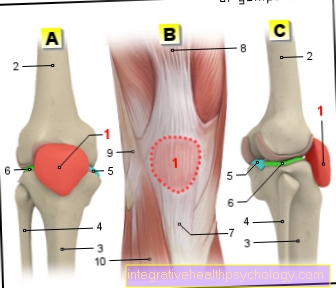
Like all joints, the knee joint is surrounded by the joint mucous membrane (synovium). It keeps the joint supple so that all movements can be carried out without friction. The infrapatellar plica is a fold of this joint mucous membrane in the knee joint. The word "infrapatellaris" indicates the position of the fold below the kneecap (patella). It is a continuation of the Hoffa fat body, which lies between the head of the tibia and the lower edge of the kneecap.

anatomy
The infrapatellar plica is a fold of the mucous membrane in the knee joint that predominantly consists of connective tissue consists. The fold of the mucous membrane is sometimes referred to as the ligamentum mucosum. It is surrounded by several layers of cells and is thus separated from the surrounding tissue. It can occur in different variants and usually separates the knee joint into two chambers. But it can also not exist at all. When it is applied, the plica is located in a bony depression below the Thighbone. From this as the intercondylar fossa near the anterior cruciate ligament marked depression pulls the mucosal fold to the anterior joint cavity and ends at the Hoffa fat body.
In its course, the plica increases in width as it runs towards the front. In some cases, the infrapatellar plica can be connected to other folds of the synovial membrane. Basically, the various folds of the mucous membrane arise during the Embryonic development and regress in the further course. At the embryo The fold of the mucous membrane acts as a kind of septum and divides the knee joint into two chambers. In adults, about 65% of the fold is formed and forms a gap at the rear edge, so that the lateral and central joint spaces are connected to one another.
course
The infrapatellar plica is located below the Kneecap. It runs through the knee joint as an extension of the fat body, which is also located under the kneecap. It divides the knee joint into a side and a middle compartment so that two chambers arise. The plica begins in a bony depression and ends at the anterior fat body. In its course it steadily increases in width. It is often in connection with the other folds of the mucous membrane, the plica suprapatellaris and mediopatellaris.
function
The infrapatellar plica and the other two folds of the mucous membrane are created in the embryonic period and often regress into adulthood. The folds of the mucous membrane do not have a direct specific function. When the infrapatellar plica is created, it forms a reinforcing fibrous cord that extends from the underside of the kneecap over the fat body into the bony depression. Occasionally it can even be too Restrictions on movement come. It can swell and block the knee joint, preventing it from sliding.
Rupture
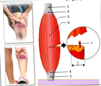
The infrapatellar plica can also tear in rare cases. Often a rupture occurs Overuse of the knee joint. Muscle weakness or an imbalance in balance can lead to tearing of various ligaments of the joint or the folds of the mucous membrane. In most cases, however, the mediopatellar plica is also affected. A rupture can be accompanied by pain, swelling of the plica and the entire joint. In MRI images is then often one Thickening of the fold of the mucous membrane to see. Depending on the severity of the injury, an operation to remove the folds of the mucous membrane may be necessary.
MRI
The Magnetic resonance imaging of the knee joint is very well suited to depict soft tissue such as muscles and organs. This also makes it possible to capture the infrapatellar plica. As a rule, it appears as a very narrow line with little signal. Poor signal means that the structure appears darker compared to the surrounding tissue. Since it is often only recognizable when one is at the same time Fluid build-up in the joint space (effusion), it is difficult to diagnose on an MRI.
OP
The various folds of the mucous membrane can be different in the knee. So it can occasionally happen that a wrinkle is thicker than normal. This can lead to friction during movement, which can also lead to entrapment. The result is not infrequently a painful inflammation that spreads to the entire knee joint and is often accompanied by swelling and an effusion. Typically, the mediopatellar plica causes this plica syndrome. If conservative therapy is not enough, surgery is often necessary. The operation takes place within the scope of a knee examination (arthroscopy), often with general anesthesia and keyhole technique.
Only small incisions are made, through which the necessary instruments with an integrated camera are pushed into the knee joint. The doctor can thus get a very good overview of the inflammatory processes in the joint. The folds of the mucous membrane as well as the inflammatory tissue are removed while protecting the articular cartilage. With this very gentle method, the patient can load his knee normally again after one to four weeks. In most cases, the operation is very successful and has few complications. The typical complaints, such as the feeling that the knee joint is blocked, usually completely disappeared afterwards. After four to six weeks, you can resume light sporting activities.
Further information also under our topic: Plica syndrome


.jpg)