Fundoscopy
Synonyms in a broader sense
Ophthalmoscopy, retinaloscopy, funduscopy, ophthalmoscopy
English: ophthalmoscopy
Definition of an ocular fundus
The fundus is the most common examination method used by an ophthalmologist. A so-called ophthalmoscope (Ophthalmoscope) illuminates the back area of the eye, i.e. the inner surface of the eye, which cannot be seen from the outside without aids. Above all, a precise assessment of the retina, vessels and the optic nerve head is possible, the changes of which can quickly provide information about certain clinical pictures.

history
The direct ophthalmoscope was developed in 1850 by Hermann von Helmholtz (*1821), who was professor of physiology and pathology in Königsberg at the time and who dealt in detail with the processes of seeing and hearing. He also invented the ophthalmometer (an instrument for determining the curvature of the cornea) in his later life.
Two years later, monocular (that is, performed with one eye) ophthalmoscopy was developed.
The further development to binocular (performed with two eyes) ophthalmoscopy did not take place until much later, around the 1950s.
Even with the indirect Ophthalmoscopy/ The fundus of the eye fixes the patient in the distance. The doctor holds a light source in one hand, which can either be an ophthalmoscope or a simple flashlight, and thus illuminates the patient's eye. With the other hand, with an outstretched arm, he places a magnifying glass at a distance of approx. 13 cm in front of the patient's eye, preferably supporting himself on the patient's forehead to enable himself to work more stable.
The image that is now visible to him is, depending on the magnifying glass, approximately 4 to 5 times enlarged, is upside down and is reversed, which is why this type of fundus mirroring requires significantly more practice to find your way. Although not so many details can be seen with this method, it does give the viewer a good overview of the retina.
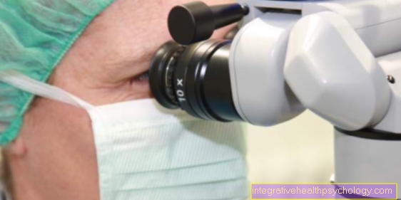
The indirect ophthalmoscopy is also binocular, i.e. with two eyes for the doctor, if the doctor carries out the examination using a slit lamp (an examination microscope) or a head ophthalmoscope. This improves the optical quality of the image it receives.
Normal findings

With a healthy one eye you don't see right in the middle, but something towards the nose shifted towards the exit of the Optic nerves (Papilla, blind spot). This is reddish to yellow, has a sharp edge, is round to oblong in shape and can have a central cavity. Here the four branches of the vessels arise from a central vessel and branch off in an arc on both sides up and down.
The arteries appear lighter and cross over the darker veins. The veins should be about 3: 2 thicker than the arteries. Further out is the yellow spot (Macula lutea), which includes the point of sharpest vision, which usually shows a yellowish color.
Complications
The fundus of the eye itself shows only a very low risk of complications. Due to the pupil-dilating eye drops there is an increased sensation of glare and the visual acuity is reduced for about three hours, which is why the patient is not allowed to drive or operate machines during this time.
In rare cases, using the eye drops may increase the pressure inside the eye, which can lead to an attack of glaucoma in those with a tendency to do so.
Implementation / procedure of the fundus examination
A fundus can only be carried out if there is a clear view of the eye. This means that there must be no clouding of the cornea or the lens of the eye or bleeding into the vitreous humor (vitreous humor).
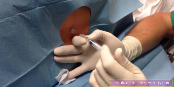
As a rule, the patient is given special pupil-dilating eye drops first to make the examination easier. The ophthalmologist then looks through the pupil at the fundus with the help of a magnifying glass. To do this, the eye must be illuminated with a bright light source. In principle, there are two methods of fundoscopy:
The direct and indirect fundus, both of which have their advantages and disadvantages.
Read more on the topic: Fundus examination
When performing a Fundoscopy the patient must fixate a point in the distance. The doctor uses his right eye to assess the patient's right eye (The same applies to the left side). For the examination he has to come very close (up to a distance of about 10 cm) to the patient, which is often perceived as uncomfortable by the patient. An electric, hand-held ophthalmoscope is brought between the eyes of the observer and the observer, which simultaneously contains a magnifying glass, a lamp and a so-called "Rekoss disk" with which the doctor and / or patient can compensate for refractive errors.
The image that the doctor sees from the fundus is approximately 15 times enlarged, upright and the right way round. Because of the high magnification, only a relatively small section of the Retina visible, but many details of the center of the retina, such as the exit of the Optic nerves, of the yellow spot or the central blood vessels.
Areas of application

Various Diseases of the eye themselves lead to specific findings on the fundus, which are Fundoscopy become visible. At a Retinal detachment if the retina and vessels lie in folds, damage to the optic nerve leads to changes in the papilla and also Pigment disorder or Tumors can thus be discovered.
At a increased intracranial pressure If the optic nerve bulges into the inside of the eye, one speaks of a congestive papilla, which is characterized by blurred edges and possible bleeding. At the glaucoma an enlarged ratio of the central cavity to the edge area can often be observed.
The fundus is also particularly important for general diseases, such as Diabetes mellitus, high blood pressure (Hypertension) or vascular calcification (arteriosclerosis). The typical finding of high blood pressure is called Fundus hypertonicus in which one can notice a pronounced constriction of the vessels and a swelling of the papillae. Bleeding is also common.
Patients with diabetes show similar vascular changes in the fundus, which, if detected early, can be reversed through better blood sugar control. For this reason, both diabetes and the other diseases mentioned above should be used at regular intervals Fundus reflections be performed.
Summary
The Fundoscopy plays a very central role, especially, but not only, in the Ophthalmology. Two examination methods are available to the doctor: the direct one Fundoscopy , which offers a strong magnification, but only a poor overview, and the indirect one Fundoscopy which provides a good overview of the retina, but does not reveal any details and only provides an inverted image.
The fundus is so often used because, firstly, it can be carried out with relatively little effort and, secondly, it allows quick conclusions to be drawn about various diseases or symptoms based on characteristic findings.