Pancreatic cancer
Note
All information given here is only of a general nature, tumor therapy always belongs in the hands of an experienced oncologist!
Synonyms
Pancreatic carcinoma (or more precise term in the narrower sense: ductal adenocarcinoma of the pancreas), pancreatic carcinoma, pancreatic cancer, pancreatic tumor
English: pancreatic carcinoma

definition
This tumor (ductal Adenocarcinoma the pancreas) is by far most common cancer of the pancreas. It belongs to the malignant neoplasms (Neoplasms).
Benign tumors (including serous cystadenoma) or other malignant forms (mucinous cystadenocarcinoma, acinar cell carcinoma) are very rare and are mentioned for the sake of completeness, but are not discussed in this topic.
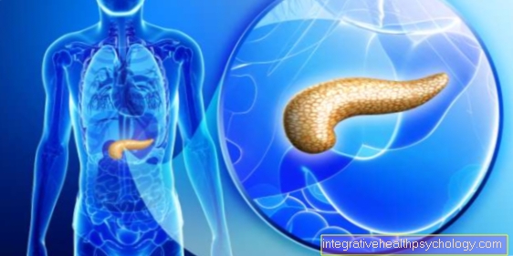
Most pancreatic cancer occurs in the front area, the so-called head of the pancreas (see anatomy of the pancreas (Pancreas)).
Epidemiology / frequency
In western industrialized nations get sick on average 10 in 100,000 Inhabitants per year. It is far more common in the USA than in Germany, Switzerland or Italy.
Sick people are usually between 65 and 85 years old. It occurs very rarely before the age of 40.
Men get sick more often than women.
causes
The exact cause of the Pancreatic cancer is unknown. However, several risk factors could be proven in the context of extensive social (epidemiological) studies.
This includes:
- long-lasting inflammation of the pancreas (chronic pancreatitis)
- To smoke cigarettes
- Alcohol abuse / alcoholism
- as well as a diet rich in fat and protein.
There are also a number of genetic diseases associated with pancreatic cancer (e.g. Peutz - Jeghers syndrome, hereditary pancreatitis, and familial pancreatic carcinoma).
Similar to other tumors of the Gastrointestinal tract the development (pathogenesis) on the basis of preliminary stages is well researched. After previous damage, new growths that do not grow in a displacing manner initially arise. These then lose more and more the similarity with their original tissue and begin to grow into the entire organ or even to cross the organ borders. The development of malignant tumors from preliminary stages through benign forms to the destructively spreading tumors is known as the adenoma-carcinoma sequence.
Illustration pancreatic cancer

Pancreatic cancer -
(Pancreatic cancer)
- Liver - Hepar
- Pancreas - Pancreas
- bile duct -
Common bile duct - Head of the pancreas -
Caput pancreatis - Pancreatic tumor (malignant)
ductal adenocarcinoma - Body of
Pancreas -
Corpus pancreatis - Pancreatic duct
( execution course) -
Pancreatic duct - Tail of
Pancreas -
Cauda pancreatisauda - Spleen - Sink
- Stomach - Guest
Risk factors:
A. - Chronic pancreatitis -
To smoke cigarettes -
Alcoholism - very fatty
and protein-rich diet
Signs / Symptoms:
B - yellowing of the conjunctiva -
Yellowing of the skin -
Lightening of the stool -
Dark urine -
Upper abdominal pain
Therapy:
C - Operation (in advance
MRI, ultrasound) -
radiotherapy
(in combination with chemotherapy) -
chemotherapy
(Drugs-cytostatics)
You can find an overview of all Dr-Gumpert images at: medical illustrations
Signs of pancreatic cancer
Pancreatic cancer signs or symptoms are difficult to pinpoint.
To make matters worse, the symptoms only develop in advanced pancreatic cancer.
Most patients are symptom-free at the onset of the disease. In this case, the disease is merely through Routine checkups (Ultrasonic etc.).
At advanced tumor involvement In the pancreas, the tumor begins to compress the duct of the pancreas, resulting in a Disturbance of the drainage of the bile fluids can be socialized. This usually results in a Yellowing of the skin and the Conjunctiva affected patients, and this usually makes them go to the doctor.
Also due to the drainage disorder of the pancreas it usually comes to one Lightening of the stool and one Urine dark in color. In some cases it can also be a so-called Fat stool come.
The combination of both symptoms suggests that there is a problem with drainage in the pancreas, although this cannot be proven. Because drainage disorders in the pancreas are also caused by Stones and Inflammation pancreatic cancer does not necessarily have to be behind the symptoms.
Sometimes patients also report belt-like abdominal pain, such as with one Inflammation of the pancreas is available.
It is then important to differentiate between inflammation and tumor involvement in this organ.
Some patients only give one Back pain with a symptom-free stomach. Often times, back pain does not indicate a malignant disease of the pancreas, which can delay the diagnosis even further.
Since the pancreas also for them Insulin production is responsible, a tumor attack can lead to a reduced supply of vital insulin, which has the consequence that the Blood sugar increases sharply and can accordingly be measured as conspicuous. In patients who initially have no diabetes is known and who suddenly suffer from fasting blood sugar levels of 400 mg / dl and more, a disease of the pancreas should always be considered.
The likelihood increases somewhat if those affected are younger patients, where one Adult diabetes can be excluded.
Symptoms
Of the Pancreatic cancer causes discomfort only in an advanced stage. He makes himself through a painless Jaundice (Jaundice), which can be traced back to a narrowing of the bile duct (ductus choledochus): The pancreatic enzymes are released into the small intestine (duodenum) to digest food. On the way from Gallbladder and liver through the Pancreatic head This duct is compressed from the outside by the tumor growth and finally compressed completely. The bile that has formed can no longer drain away, it backs up in the gallbladder and liver and passes into the blood there. The white skin of the eyes (sclera) turns yellow.
If not treated in time, this leads to so-called mechanical Jaundice (Jaundice) (due to congestion of the bile ducts) to progressive damage to the liver so that it can no longer fulfill its extensive metabolic tasks (Hepatic insufficiency).
Illustration of the pancreas

- Body of
Pancreas -
Corpus pancreatis - Tail of
Pancreas -
Cauda pancreatisauda - Pancreatic duct
( execution course) -
Pancreatic duct - Duodenum lower part -
Duodenum, inferior pars - Head of the pancreas -
Caput pancreatis - Additional
Pancreatic duct -
Pancreatic duct
accessorius - bile duct -
Common bile duct - Gallbladder - Vesica biliaris
- Right kidney - Ren dexter
- Liver - Hepar
- Stomach - Guest
- Diaphragm - Diaphragm
- Spleen - Sink
- Jejunum - Jejunum
- Small intestine -
Intestine tenue - Colon, ascending part -
Ascending colon - Pericardium - Pericardium
You can find an overview of all Dr-Gumpert images at: medical illustrations
Other symptoms (upper abdominal and back pain)
Upper abdominal pain
Other common symptoms are dull Upper abdominal painthat can radiate into the back and cause weight loss. This is caused by a disturbance in the utilization of food (maldigestion): In addition to the bile duct, the pancreatic duct (ductus pancreaticus), which also runs within the gland, can be compressed, so that the pancreatic ducts that are required to break down the food, formed by the pancreas and released into the pancreatic duct Enzymes can no longer reach their target in the small intestine. This symptom is associated with inflammation of the pancreas and is therefore also called secondary obstructive pancreatitis (i.e. occlusive inflammation of the pancreas). Occasionally there is also a painful but not dangerous inflammation of the superficial leg veins (a so-called Thrombophlebitis) on.
Back pain
Many patients with pancreatic cancer suffer from it Back pain.
However, the reverse of this sentence hardly applies. More than every second person has back pain in their lifetime. This makes this clinical picture one of the most common. The causes are often tension in the muscles or spinal disorders. Because back pain is so common, doctors rarely associate it with pancreatic cancer. They are a very unspecific symptom. Anyone who goes to the doctor with back pain should not fear a cancer diagnosis. Back pain often goes away with time or with the administration of pain relieving medication such as Diclofenac.
The cause of pancreatic cancer-related back pain is probably due to the irritation from surrounding annoy. It is not uncommon for tumor diseases to be coupled with inflammation of the surrounding tissue. Since the tail of the pancreas in particular is very far back in the abdomen, a tumor in this area can cause back pain.
histology
Tumors of the pancreas originate in the duct epithelia, i.e. surface cells of the pancreatic ducts (see anatomy). The tumor forms glandular structures that can also produce mucus and spreads diffusely throughout the organ.
Because the pancreas is not surrounded by a protective, connective tissue capsule, the tumor quickly grows beyond the pancreas and affects surrounding structures. This explains the extremely poor prognosis for pancreatic cancer (see below). The risk of settlements (metastases) that are carried by the bloodstream to other organs is high, as the tumor tends to grow into blood vessels.
Resettlements are particularly common in
- the liver (65%)
- the lungs (25%)
- and in the skeleton (10%)
to find.
Diagnosis of pancreatic cancer

If the symptoms described are suspected, a tissue sample is taken from the pancreas (fine needle aspiration). The atypical appearance of the tumor cells allows them to be clearly identified under the microscope (histopathological diagnosis). Particular caution is required when taking samples, as there is a risk that the tissue does not come from the tumor but accidentally from the surrounding, “only” inflammatory part of the organ. Primarily, however, the long symptom-free course, not a false detection, is responsible for a late diagnosis of pancreatic cancer.
Other diagnostic options are:
- an ultrasound examination (sonography)
- Computed tomography, CT
- MRI, magnetic resonance imaging
- Tumor markers
Proteins produced by the tumor can be detected in the blood. (So-called tumor markers, these are somewhat specific for a certain tumor: those used especially in pancreatic cancer are called CEA and CA 19-9. Finding new markers for different tumors is the subject of current research.).
Changes in the blood count in pancreatic cancer
In addition to a physical examination and ultrasound and CT examination, the diagnosis of pancreatic cancer also includes an examination of the blood.
Although there are no specific values that are only changed in the blood in the case of pancreatic cancer, there are some blood values that can generally indicate a malignant disease.
To be mentioned
- high blood sugar levels with diabetes mellitus not described above
- high inflammation values (CRP value, leukocytes)
- as well as elevated pancreatic enzymes (e.g. lipase)
Read more on the topic: Lipase increased and lipase in the blood - what does it say ?.
An iron utilization disorder with low iron values in connection with reduced hemoglobin values in the blood can also indicate tumor-related anemia.
forecast
The prognosis is very bad. By late diagnosis (the tumors are then usually already a few centimeters) are only 10 to 15% operable. Few patients survive longer than 5 years.
The pancreatic cancer speaks bad on chemotherapy on. This is why the mortality rate is high with a comparatively low incidence of the disease (around 3% of all cancers): this tumor is the fifth most common of all cancer deaths.
Chances of survival
Basically, unfortunately, it can be said that the chances of survival of a pancreatic cancer (pancreatic cancer) regardless of type and spread extremly bad are.
One reason for this is the anatomical location of pancreasshowing tumor growth in almost all Upper abdominal organs and on the other hand the diagnosis that is often made very late. Symptoms only develop at an advanced stage and a preventive examination for pancreatic cancer is not the standard in Germany. Therefore, the disease is recognized late. The method of choice for diagnosing pancreatic cancer is the Ultrasonic of the upper abdomen and, if suspected, one Biopsy (tissue sample) with pathological findings.
The only curative therapy for pancreatic cancer is that surgical removal. However, this can only take place completely if the tumor has spread only slightly.
Overall are only around 20 % the tumors are completely resectable. Even after such an intervention, the patient's likelihood is further 5 years to survive only at about 10 %. In the remaining patients with inoperable pancreatic cancer, this probability is below 1 %.
Often they die after about 10 months as a result of their cancer. This number varies slightly between individual patients depending on their overall wellbeing and response to chemotherapy and radiation therapy.
Overall, pancreatic cancer is one of the cancers with the worst prognosis.
Terminal stage
Because the diagnosis is usually very late, the disease often progresses quickly with and without treatment.
Although there are ways to curb the progress of tumor growth, this is usually the case no cure more achieved. First it tries to drain the Bile acids restore. This is usually achieved by inserting a Stents into the pancreatic duct.
It is also possible to try to remove the part of the pancreas affected by the tumor, sometimes with very complex operations. Also one chemotherapeutic measure can be used.
therapy

There are essentially three different therapy options available for treating pancreatic cancer:
- surgery
- radiotherapy
- chemotherapy
Treatment can be done by just one option or by a combination. The choice of therapy depends on many different factors. This includes, for example, the age and gender of the patient, but also the operability of the tumor and the progression of the disease. The final therapy of the tumor is often determined by doctors from various disciplines in the course of a so-called tumor conference. Internists, surgeons, pathologists, anesthetists, etc. are involved.
The method of choice for treating pancreatic cancer is surgery. It is the only curative option to completely remove the tumor. In advance, the extent of the tumor should be recorded using various imaging methods (e.g. MRI, ultrasound, etc.).
In addition, lymph node involvement and any distant metastases should be clarified by the doctor. All of this is important to be able to assess the operability of the cancer.
In principle, the following applies to all tumor diseases: the lower the spread, the better the chances of treatment. But what happens during such an operation? The type of intervention depends on the location of the tumor. If it is in the tail of the pancreas, either only this part is removed or, if the extent is unclear, the whole gland. Usually the spleen must also be removed.
The aim of the operation is to remove the tumor as completely as possible. To do this, part of the surrounding, healthy tissue must also be cut out. In this way, one tries to prevent individual cancer cells that may have broken off from the tumor from being resected. Often, immediately after removal, the tumor border is examined under the light microscope to determine whether the resection border is tumor-free. In this case one speaks of an R0 resection.
Much more often, however, the pancreatic cancer is located in the area of the head of the organ. This lies directly on the duodenum and the stomach. In this case, the so-called Whipple operation is used, which is much more complicated than resection of the pancreas tail.
Whipple involves removing the head of the pancreas or the entire pancreas, the duodenum, bile duct with gallbladder, surrounding lymph nodes and parts of the stomach. This is necessary because the pancreas is close to these organs and there is therefore a risk of the tumor spreading.
It is hoped that this extensive procedure will completely remove all tumor cells. Since the connection between the stomach and small intestine is missing after the removal of the duodenum, the digestive tract has to be surgically reconstructed. To do this, a loop of the small intestine is sutured to the stomach and, depending on the extent of the resection, a connection is established between the pancreatic outlet and the biliary system. This ensures that the patient's digestion works reasonably well after surgery.
Often times, patients find it difficult to digest after Whipple surgery. As a result, a diet should be followed and the portion size reduced.
If the pancreas is completely removed, hormones such as insulin must also be replaced with medication.
If it is not possible to remove the entire tumor during the operation, its size must be reduced either before or after the operation using radiation and chemotherapy. In this case one speaks of a neo- or adjuvant therapy.
A second major branch of the therapy for pancreatic cancer in addition to surgery is chemotherapy. Chemotherapy is the treatment of a tumor using drugs that inhibit the growth of cells. These drugs are known as cytostatics. They work especially well on cells that are growing rapidly and dividing quickly. In addition to tumor cells, these criteria are also met by healthy cells such as hair root cells or the cells of the blood-forming bone marrow. This results in the well-known side effects of chemotherapy (hair loss, paleness, tendency to bleed, nausea, etc.). Basically, however, it should be noted that pancreatic cancer can never be cured by chemotherapy alone. This always requires an operation. Capecetabine and Erlotinib are a few examples of cytotoxic drugs that are used to treat pancreatic cancer.
Chemotherapy often consists of a cocktail of different cytostatics with different mechanisms of action. It is hoped that a lower dosage of individual active ingredients will result in fewer side effects.
The last major curative therapy option is radiation therapy. This often happens before or after the operation in combination with chemotherapy. Radiation therapy is a targeted attempt to destroy cancer cells with the help of radioactive radiation. Thanks to modern technology, the radiation dose can be concentrated almost exclusively on the tumor tissue, so that as little healthy tissue as possible is damaged. As with chemotherapy, radiation therapy alone cannot cure pancreatic cancer. This is still not possible today without an operation.
If the tumor cannot be cured, palliative medical treatment must be used. Palliative measures are therapies that do not cure the tumor, but rather keep its effects as low as possible. The aim of palliative medicine is to make the rest of the patient's life as comfortable as possible. The focus here is on pain therapy. For most patients, tumor pain is the greatest impairment. Treatment is carried out using common oral pain relievers such as paracetamol, but in more severe cases also intravenously, for example with morphine.
Read more on the subject at: Palliative medicine
In addition to combating pain, attempts are also made to treat the classic symptoms of pancreatic cancer. For example, jaundice, which is caused by an obstruction of the bile duct due to strong tumor growth, can be treated in a minimally invasive manner using a stent. In the case of severe nausea and vomiting, the option of a gastric tube must be considered.
You can find more information about the treatment of pancreatic cancer at:
- Therapy pancreatic cancer
Age
In principle, everyone can use a pancreas get cancer at any age. Nevertheless, the risk of illness increases sharply with age.
Pancreatic cancer in children is very rare and rarely observed. In old age 50 years he can sporadically occur, but primarily in patients exposed to many risk factors.
The main risk factors are Smoke, Diabetes mellitus Type 2, chronic pancreatitis e.g. by Alcohol abuse and Obesity.
A familial accumulation of the disease is also discussed. In addition, men are said to get sick more often than women, presumably because of increased alcohol and nicotine consumption.
With regard to the frequency of the disease, the zenith is in the region of 70 years.
Further subject areas
Further information on this topic can be found at:
- Pancreatic Cancer Age
- Pancreatic cancer signs
- Pancreatic Cancer Causes
- End-stage pancreatic cancer
- Pancreatic cancer prognosis
- Pancreatic cancer therapy
- Pancreatic cancer and back pain
- metastasis
- Water in the stomach
- Verner-Morrison Syndrome
All topics that have been published on the field of internal medicine can be found at: Internal medicine A-Z

