The calcified kidney
What is a calcified kidney?
The calcified kidney (also called nephrocalcinosis) is a disease in which more calcium is deposited in the kidneys. The causes can be very different, but mostly a metabolic disorder is the cause. The consequences are kidney dysfunction up to complete kidney failure.
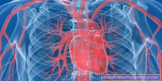
Occasionally, a calcified kidney is also used to refer to the calcification of the renal artery, i.e. the vessel that supplies the kidney with blood. In this case, kidney function may also be impaired. The causes of the disease, however, are more likely to be found in cardiovascular diseases, i.e. in calcium and fat deposits in the vessels.

The causes of a calcified kidney
The causes of a calcified kidney are usually a disturbed calcium metabolism. For example, increased absorption in the intestine can lead to more calcium deposits in the kidneys. The bone metabolism can also produce more calcium than usual and thus lead to an accumulation of calcium.
In most cases, an existing impairment of kidney function is also involved in the development of the disease. Due to the reduced kidney function, the calcium is no longer excreted sufficiently, instead it accumulates in the kidney. This in turn worsens kidney function, which can create a vicious circle.
The deposits can also occur in the context of other diseases such as storage diseases or tumor diseases. This changes the way calcium is processed in the body, which may lead to calcium deposits. In addition, congenital kidney diseases can lead to impaired kidney function, which can occur even before birth. As a result, kidney calcifications occur even in children.
Calcification of the kidney can also occur in the form of kidney stones, in which case the calcification collects in one place and forms a stone in the kidney tissue.
Find out all about the topic here: Renal impairment.
Kidney stones
Kidney stones are the accumulation of calcium deposits in a specific place so that so-called concretions arise there. The reason for this is often in too little drinking combined with a diet that is rich in oxalate (e.g. in spinach). Recurring urinary tract infections, some of which reach the kidneys, also promote the development of kidney stones. If metabolic diseases occur that increase the excretion of calcium in the urine or if this excretion is disturbed, a lot of calcium accumulates in the kidneys. This can also lead to the development of kidney stones.
In the case of kidney stones, familial accumulations have also been reported, which is why it is assumed that the disease is a genetic component. Usually, the stones are initially invisible. Symptoms only appear when the stone comes off and gets stuck in the ureter or when it obstructs the entrance to the urinary tract in the renal pelvis. The result is colic-like pain, sometimes what is known as hematuria, in which blood cells get into the urine and the urine turns red as a result.
The disease can best be diagnosed on ultrasound. There the stones stand out as a lightening in the kidney tissue. The kidney stones can also be detected in other imaging such as X-rays or CT.
Therapy consists of removing the kidney stones. This can be done by means of an operation or also through shock wave therapy. Subsequently, affected people should ensure that they drink sufficient quantities, and there are also drugs that improve calcium excretion, which means that less calcium is absorbed in the kidney tissue.
Find out all about the topic here: Kidney stones.
The symptoms of a calcified kidney
A calcified kidney is often an incidental finding, as initially little or no symptoms occur. Only when the disease is well advanced can the first symptoms be noticed.
The calcification of the kidneys mainly leads to disturbances in excretion. For example, more proteins can get into the urine, which sometimes leads to foamy urine. Admixtures of other cells, such as red blood cells, can also be seen in the urine.
If kidney function is severely restricted, water retention occurs, especially in the legs. This suggests that the kidneys are no longer excreting enough water.
You can find more information on this topic here: Symptoms of renal insufficiency.
Pain in the calcified kidney
A calcified kidney does not usually cause any pain at first. However, if calcium is deposited in the form of real kidney stones, these can prevent the urine from flowing out. Urine accumulates in the kidneys, which can be felt as pain.
Typically, stones do not appear on both sides at the same time, so that the pain can only be felt on one side. Those affected usually complain of flank pain.
The diagnosis of the calcified kidney
The diagnosis of the calcified kidney is best made on ultrasound. Calcifications in the tissue can be seen there particularly well. A blood test can also provide evidence of kidney calcification. On the one hand, the kidney function values can be determined there. If these are decreased, this indicates decreased kidney function. Typically, the creatinine is increased and the GFR (filtration rate of kidney corpuscles) decreased. An increased level of calcium in the blood can also sometimes be seen.
The urine should also be examined. If this is particularly acidic, this also indicates limescale deposits. The calcification can destroy the kidney corpuscles, which can lead to an increased excretion of protein and cells. These can also be determined in the urine test using the U-Stix.
Read more about the topic here: The urine test.
What do you see in the ultrasound?
Calcified kidneys can produce very different images on ultrasound. If, for example, a kidney stone appears, the ultrasound shows a clear brightening of the stone. This phenomenon is usually one-sided and cannot be observed on both kidneys at the same time.
In diseases that affect the entire body (e.g. metabolic diseases), both kidneys are usually equally affected. Lime splashes in the kidneys (many small white spots) or a general lightening of the kidney tissue can be observed.
Also read the article: Ultrasound of the abdomen.
Therapy of the calcified kidney
Therapy of the calcified kidney is initially conservative (treatment that takes place through medication or physiotherapy) and is directed against the underlying disease that caused the calcification. If the reason is too high a calcium level, a low-calcium diet should be observed. There are also drugs that cause more calcium to be excreted. This prevents it from being deposited in the kidney.
Conservative therapy options also include increased intake of fluids. Due to the increased general excretion, more calcium can also be dissolved in the urine and thus transported out of the body. Another disease that can cause a calcified kidney is renal tubular acidosis, which leads to functional disorders in the kidneys and thus to an incorrect elimination of electrolytes. Depending on the type of renal tubular acidosis, different drugs are used that lead to an increased or decreased sodium excretion or an altered potassium excretion. Diuretics (water tablets) can also be taken.
Proper nutrition
In the case of the calcified kidney, a reduced intake of calcium should be ensured in the diet. Since calcium is mainly found in dairy products, milk, yoghurt, quark, pudding and cheese should be avoided if possible.
In addition, you should not consume foods that contain relevant amounts of oxalate. The oxalate forms a complex with the calcium in the kidneys and thus promotes the development of kidney stones. Oxalate is found in blueberries, beetroot, spinach, Swiss chard, parsley, etc.
Find out more about this topic at: Diet for urinary stones.
When do I need an operation?
An operation is usually sought if conservative therapy options do not bring the desired effect. If, for example, kidney stones also appear in the calcified kidney, these should be surgically removed.
The operation is usually very small because it can be performed with instruments that can be pushed along the entire urinary tract. Often only a small or no abdominal incision is necessary.
An operation can also be useful if the underlying disease can be treated surgically.This is the case, for example, with the malfunction of the parathyroid gland. If this leads to an increased supply of calcium in the body, removing the parathyroid glands can improve the calcified kidney.
These operational options exist
In the case of the calcified kidney, a distinction must be made between various surgical options that are used depending on the severity of the disease. If kidney stones have already formed, they can be removed endoscopically, i.e. using a device on a long tube. The stones can also be destroyed by means of shock wave therapy; the stone fragments may then also have to be retrieved from the kidney endoscopically. Open surgery is rarely necessary for stones.
If the kidney is particularly heavily calcified, it sometimes happens that its function is so restricted that part or all of the kidney has to be removed. One would like to avoid such an operation if possible through other therapeutic measures.
For more information, see: Therapy of kidney stones.
The course of the disease of the calcified kidney
Calcified kidney disease progresses without treatment for the disease. While initially only small calcifications are deposited, this increases over time. The kidney therefore initially appears, for example in ultrasound, with only slightly lightened tissue. Gradually, however, the calcium deposits compact until there are many small calcium splashes or one or more kidney stones develop.
At the same time, the kidney function is worsened by the calcium deposits, so that waste products of the metabolism are more poorly excreted, fluid is also no longer sufficiently excreted at some point and water retention occurs as a result of this renal insufficiency.

