Gallbladder cancer
Synonyms in a broader sense
Gallbladder tumor, gallbladder carcinoma, squamous cell carcinoma, adenocarcinoma, porcelain gallbladder
Note
All information given here is only of a general nature, tumor therapy always belongs in the hands of an experienced oncologist (tumor specialist)!
definition
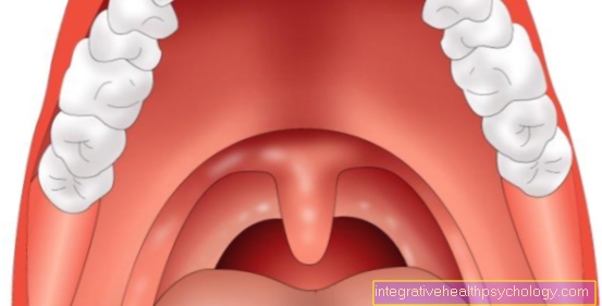
Gallbladder cancer (gallbladder cancer) is a rare but very malignant tumor with a poor prognosis because the symptoms are as painless Jaundice (Jaundice) often attract attention late. A distinction is made between two different types of tumors. The Squamous cell carcinomawhich is particularly malicious and that Adenocarcinomawhich occurs more often. The disease comes predominantly after 60 years of age and affects women twice as often as men. A long time Gallstone disease and chronic Gallbladder infections are considered risk factors for developing gallbladder cancer.

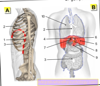
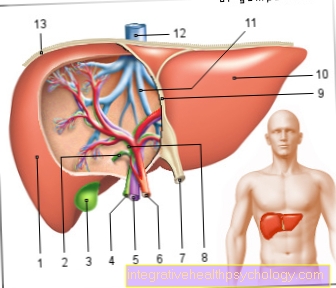
Illustration of the gallbladder

- Gallbladder Body -
Corpus vesicae biliaris - Right liver bile duct -
Ductus hepaticus dexter - Left liver bile duct -
Left hepatic duct - Gallbladder duct -
Cystic duct - Gallbladder Neck -
Collum vesicae biliaris - Mucous membrane -Tunica musoca
- Common
Liver bile duct -
Common hepatic duct - bile duct -
Common bile duct - Pancreatic duct -
Pancreatic duct - Extension of the united
Execution corridor -
Ampula hepatopancreatica - Large duodenal papilla -
Major duodenal papilla - Duodenum Descending Part -
Duodenum, descending part - Liver, diaphragmatic side -
Hepar, Facies diaphragmatica - Pancreas -
Pancreas
You can find an overview of all Dr-Gumpert images at: medical illustrations
frequency
Gallbladder cancers are very rare and affect only about 1 in 100,000 residents. However, gallbladder cancer is 3 to 5 times more common than bile duct cancer. The sick are mostly older than 60 years and women are affected twice as often.
Tumor types
Two types of cancer can develop in the gallbladder wall. First of all, the less common squamous cell carcinoma, which arises from the surface cells (epithelial cells) of the gallbladder mucosa and is characterized by a particular malignancy. The more common adenocarcinoma, arises from gland cells in the gallbladder lining and is slightly less malignant than squamous cell carcinoma.
Causes and Risk Factors

Long-term inflammation of the gallbladder is one of the risk factors for the development of gallbladder cancer (chronic cholecystitis).
Besides, that seems Gallstone disease (Cholecystolithiasis) to play a role. Because 80% of that cancer Sufferers also have gallstones in the Gallbladder, but by far not every patient (only approx. 1%) with one Gallstone gets a gallbladder carcinoma.
Approx. 3-5% of patients will after one Salmonella infection to so-called Permanent eliminators. This means that the bacteria could not be completely killed and the patient always excretes salmonella in his stool. At the same time, the gallbladder is colonized with salmonella, which is also a risk factor for gallbladder carcinoma.
Long-standing chronic inflammation of the gallbladder can lead to one Calcium deposits (calcification) on the inner wall of the gallbladder come. This state is also called Porcelain gall bladderwhich are precancerous (Precarcinosis) for gallbladder cancer (gallbladder cancer).
Benign tumors the gallbladder (Gallbladder adenomas) should be removed from a size of 10 mm, because they have a certain potential for malignant degeneration. Adenomas smaller than 10 mm should be checked sonographically every six months. Occasionally also occur in the gallbladder Gallbladder polyps which, however, have little potential for degeneration.
Symptoms
Most of the time there are no symptoms in the early stages, which is why the disease only attracts attention at an advanced stage. The first symptom is usually one painless jaundice (Jaundice), which is caused by the narrowing of the bile ducts by the tumor, causing it to build up bile in the liver comes. The symptoms of jaundice are one Yellowing of the skin and the white eye color (Dermis, sclera) and an onerous itching as a result of bile salts deposited in the skin. There is also a clay-like one Discoloration of the stool, by the lack of the bile pigment and a Urine dark in colorbecause the kidney takes over the excretion of the bile pigments. Due to the lack of the bile acids in the Small intestine fats can be digested more poorly, leading to intolerance to high-fat meals and too Fatty stools (Steatorrhea) can come. Very rarely it can too Pain in the right arm come as the pain projects into this area as the gallbladder is here her "Dermatome" has, i.e. the area in which we feel pain when something is wrong with the organ.
If the outflow from the gallbladder is prevented, a bulging gallbladder under the right costal arch can be felt in addition to the painless jaundice. This symptom complex is also called Courvoisier's symbol designated. Other complaints can be unspecific, diffuse Upper abdominal pain, Nausea, vomiting, loss of appetite, and indigestion. Pain in the right upper abdomen and other unspecific symptoms that can occur with most cancers, such as weight loss (tumor cachexia), anemia, fatigue and listlessness, can be added as late signs.
-> Read on on the topic Gallbladder Cancer Diagnosis
Tumor spread (metastasis)
You can do different Forms of metastasis describe:
- Lymphogenic metastasis
The lymph vessels drain the Lymph fluid from all parts of our body, especially the gallbladder, has an excellent lymph supply. If the tumor is connected to a lymphatic vessel as it grows, it can easily happen that some cells become detached from the tumor cell cluster and are carried away with the lymph flow. There are numerous lymph nodes in the course of a lymphatic vessel. In them is the seat of the immune defense, which has the task of catching germs (bacteria) and fighting them. The tumor cells settle in the nearest lymph nodes and multiply again there. This creates a lymph node metastasis. In this type of cancer, lymph nodes that are in the immediate vicinity and later also those in the course of the main artery (aorta) are affected. This type of cancer is characterized by very rapid lymphogenic metastasis, so that it is always advisable to also remove the surrounding lymph nodes during the surgical bile removal.
- Hematogenous metastasis
If the tumor gains connection to a blood vessel as a result of its growth, cells can tear themselves loose in this situation as well and be dispersed throughout the body via the bloodstream (hematogenous). As the first station, the blood flows through the liver, where the carcinoma cells can settle and form daughter ulcers (distant metastases). In the further course of the disease, cells can also detach from the liver metastases and spread further into the lungs. Later it can too Metastasis come into the peritoneum, which is also called peritoneal carcinosis, and into the ovaries, the skeletal system or the spleen.
- Per continuitatem
The tumor can grow into other neighboring organs as it spreads (tumorous infiltration). Not infrequently this is the case with gallbladder cancer (gallbladder cancer) at the time of diagnosis. For example, gallbladder cancer can spread to the liver Duodenum (Duodenum), grow into the pancreas (pancreas) and other adjacent structures.
Staging
However, an exact assessment of the tumor stage is often only possible after the operation, when the tumor has been removed and the surgical specimen (resected material) and the lymph nodes have been examined with a microscope (histologically).
T stages:
T1: Infiltration of the mucous membrane (mucosa) or muscles
- T1a: Mucosal infiltration
- T1b: Infiltration of the muscles
T2: Infiltration of the connective tissue (serosa) following the muscle layer
T3: Perforation of the last organ-encasing layer (serosa, visceral peritoneum) and / or ingrowth (infiltration) into the liver or other neighboring organs (e.g. duodenum, stomach, bile ducts).
T4: Infiltration of the portal vein (Vena portae) or hepatic artery (Arteria hepatica)
or infiltration of 2 or more neighboring organs
N stages:
N0: No lymph node metastases detectable
N1: Surrounding (regional) lymph node metastases between the porta hepatica and the duodenum (hepatoduodenal ligament) affected
N2: Other nearby lymph node metastases
M stages:
M0: no distant metastases detectable
M1. Distant metastases (especially liver, later also lungs)