Yellow spot
Synonyms
Medical: Macula Lutea (Latin)
English: macula
definition
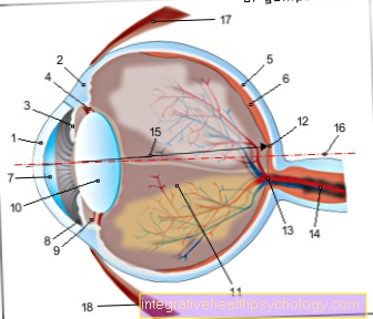
Yellow spot is a circular area on the retina of the eye where the highest density of photoreceptor cells is present.
If you fix an object with your eyes, the light of this object is thrown through the lens of the eye exactly into the area of the yellow spot. The eponymous color of the yellow spot is due to the high concentration of different, carotene-like pigments (which also give the carrot its color) in the retina. When looking at the fundus through an eye fundus reflection, however, the yellow spot hardly appears noticeable.

construction
The yellow spot has a size of about 5mm and can be further found in the visual pit (lat. Fovea centralis), Parafovea (para = next to, adjacent) and Perifovea (peri = around something).
The pit of vision, which is located in the middle of the yellow spot, is the place of sharpest vision. It only contains cones that are responsible for color vision. It closes to the outside of the approximately 0.5 mm wide Parafovea in which the proportion of rods increases. Rods are important for night vision because of their high sensitivity to light, but they cannot distinguish between colors. The highest density of rods is found in the outer area of the yellow spot, in the Perifovea - an area that takes up the outer 1.5mm.
Read more about this: Anatomy of the eye
Function of the yellow spot
The high concentration of cones in the central area of the yellow spot results in the high resolution of our central field of vision, which is a prerequisite for reading, for example.
Everyone can easily determine for themselves that the resolving power of the rest of the retina is insufficient for this by concentrating on not making saccade movements while reading, i.e. not jumping from one word to the next. If you focus your gaze on one word, it is extremely difficult to decipher neighboring words.
However, since cones are not light-sensitive enough to see in the dark, the high resolution of the central area of the yellow spot is lost, e.g. at night, and we mainly see with the rods of the Peri- and Parafovea, i.e. the edge areas in the yellow spot. A fact that everyone can easily check by trying to fix their eyes on a very weakly shining star in the sky. The light appears clearer if you look past the star you are looking at.
The fact that we do not notice this division of tasks and the limitations of the various areas of our field of vision is due to the ability of our brain to create a stable image from various impressions through many eye movements.
You may also be interested in the following topics:
- How does seeing work?
- Visual acuity

- Cornea - Cornea
- Dermis - Sclera
- Iris - iris
- Radiant Bodies - Corpus ciliary
- Choroid - Choroid
- Retina - retina
- Anterior chamber of the eye -
Camera anterior - Chamber angle -
Angulus irodocomealis - Posterior chamber of the eye -
Camera posterior - Eye lens - Lens
- Vitreous - Corpus vitreum
- Yellow spot - Macula lutea
- Blind spot -
Discus nervi optici - Optic nerve (2nd cranial nerve) -
Optic nerve - line of sight - Axis opticus
- Axis of the eyeball - Axis bulbi
- Lateral rectus eye muscle -
Lateral rectus muscle - Inner rectus eye muscle -
Medial rectus muscle
You can find an overview of all Dr-Gumpert images at: medical illustrations
What is the difference between the yellow and the blind spot?
The yellow spot is the point of sharpest vision, as this is where the highest density of color-sensitive light receptors is found on the retina. It lies exactly in the visual axis. An image that is in the center of the field of vision falls on the yellow spot. Next to it in the direction of the nose is the so-called blind spot. This is the point at which the optic nerve reaches the eye. In addition, several vessels enter the eye from here. Therefore, light receptors are missing at this point.
While the yellow spot is the point of sharpest vision, the eye completely lacks visual information at the blind spot. However, the brain completely compensates for this with the second eye.
Further information on this: Blind spot
Yellow spot diseases
The most important disease of the yellow spot is macular degeneration, which affects around 2 million people in Germany alone.
This leads to the death of the sensory cells and thus to the partial blindness of the person concerned. The causes for this can be varied: The majority of those affected suffer from age-related macular degeneration (AMD). In addition to age, smoking and various genetic predispositions are the main causes.
Macular degeneration can also occur with severe myopia or the side effects of various drugs (certain anti-rheumatic drugs and preventive drugs against malaria).
Below is an overview of the most common and relevant diseases of the yellow spot.
Macular degeneration
In macular degeneration, the yellow spot deteriorates slowly. Since this is the point in the center of the field of vision, the central visual acuity decreases until it finally comes to blindness.
The most common form is senile, age-related macular degeneration. A distinction is also made between wet and dry macular degeneration.
In wet macular degeneration, new blood vessels form in the area of the yellow spot. However, these vessels are of poor quality, so bleeding can easily occur. In the much more common dry macular degeneration, these new vessel formations are absent. It runs much slower.
You can find more about this clinical picture on our website: Macular degeneration
Macular edema
Macular edema is an accumulation of fluid in the area of the yellow spot. It occurs, for example, with inflammation of the retina or choroid. Vascular diseases, for example due to diabetes mellitus, can also lead to macular edema. The yellow spot can swell due to the accumulation of fluid and the field of vision appears blurred.
Further information on this: Choroidal inflammation
Macular ectopy
A displacement of the yellow spot from the central visual axis is called macular ectopy. As a result, an image in the center of the field of view no longer necessarily falls on the yellow spot, which can impair vision.
This can be congenital, or it can result from an illness or an operation. In congenital macular ectopia, the brain can bring the shifted yellow spot back into the center of the visual axis by squinting; this is known as pseudostrabism.
Who discovered the yellow spot?
The yellow spot was discovered by Samuel Thomas von Soemmering, a German anatomist.


