Bone chipping
General
Bridges can occur more or less frequently on almost every bone in the body. These can be caused by injuries or signs of fatigue. Certain diseases can also increase the risk of bone fractures. In most cases, an external force on the bone is what causes a bone fracture. How the bone breaks in the end depends on many factors. The amount of force applied and the shape of the object that acts on the bone are particularly important. For example, a bone fracture that occurs while exercising typically takes on a different shape than a bone fracture that is diagnosed after a serious automobile accident.
Depending on the individual situation, the affected bone can break into different numbers of parts. At a large variety of parts one also speaks of one Comminuted fracture. These are particularly common after blunt and widespread violence on the bones. When a small piece of bone separated from the main fragment was is from one Bone chipping the speech. These occur mainly in connection with fractures. A bone splinter can cause problems due to its unfavorable location and should be treated differently depending on its individual location. Individual advice on therapy options in the case of a bone fragment is best provided by one Specialist in Orthopaedics as they are considered experts in the field of bone fractures and chipped bones.
Symptoms of bone chipping
The symptoms of a bone fragment can vary widely. So the type and intensity of the symptoms mainly depends on the person Size of the splinter, the compromised bones, as well as the Localization of the piece of bone from. Often, symptoms of the bone fragment are overshadowed by other symptoms caused by the underlying injury. This can mean that small fragments of bone are initially not discovered. Especially with simultaneous fractures that affect the entire bone strong pain in the foreground. It can also be in the area of injury to one bruise as well as one swelling come to the skin. Depending on which bone the chipping belongs to, can Restrictions on movement and Pain when moving occur. Particularly in places where nerves and blood vessels run close to the bone, side effects such as Loss of sensitivity, which can arise on the one hand from damage to the nerve from the underlying injury, but also from direct impairment from the bone splinter. In rare cases it can be due to the bone splinter too Injuries to the vessels come with profuse bleeding as a result.
Appointment with ?

I would be happy to advise you!
Who am I?
My name is I am a specialist in orthopedics and the founder of .
Various television programs and print media report regularly about my work. On HR television you can see me every 6 weeks live on "Hallo Hessen".
But now enough is indicated ;-)
In order to be able to treat successfully in orthopedics, a thorough examination, diagnosis and a medical history are required.
In our very economic world in particular, there is too little time to thoroughly grasp the complex diseases of orthopedics and thus initiate targeted treatment.
I don't want to join the ranks of "quick knife pullers".
The aim of any treatment is treatment without surgery.
Which therapy achieves the best results in the long term can only be determined after looking at all of the information (Examination, X-ray, ultrasound, MRI, etc.) be assessed.
You will find me:
- - orthopedic surgeons
14
You can make an appointment here.
Unfortunately, it is currently only possible to make an appointment with private health insurers. I hope for your understanding!
For more information about myself, see - Orthopedists.
causes
The causes for the development of bone chipping are very diverse. What they all have in common is that a more or less strong force acts on the bones and turns them into one Detachment of the bone part leads. In general, a distinction must be made between two different forms of bone fragmentation. On the one hand, bone splintering can be caused by a Force the affected bone is otherwise intact. On the other hand, bone splinters are often found in the context of complete bone fractures. In most cases, bone splintering occurs on the basis of a complete bone fracture.
At a Debris break In addition to larger bone fragments, small bone fragments can also occur. But also with other bone fractures in which there are only two other bone fragments in addition to the bone splinter, splintering can occur.
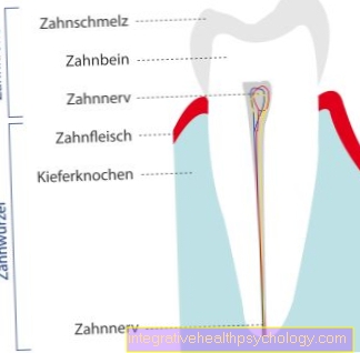
Reasons for the development of a bone chipping without a complete bone fracture are numerous and very different from person to person. So it can be next to one external violence Injuries can also occur, which are secondary to bone chipping. For example, if a Bone pops out of the jointthis can cause chipping. This is often the case with a so-called dislocated upper arm for example the case.
In addition, ligament tears can occur, in which a piece of the bone to which the ligament was previously attached also tears out. This is particularly common with ligament injuries to the hand. For example, bone splintering can occur if the collateral ligament of the thumb is torn out in an accident. The injury often occurs in skiing and is therefore also known as the skier's thumb.
therapy
The individual therapy for an existing bone fragment is very different. In principle, this depends on the structures involved and the size of the splintering. In general, a distinction can be made between surgical and conservative therapy.
In addition to taking pain medication, conservative therapy usually includes stabilizing the affected bone. How strong the stabilization should be depends on the size of the fragment and the affected area of the body. Bones that are heavily stressed in everyday life should usually receive strong stabilization. External stabilization can be ensured by applying a plaster of paris or wearing a splint. A plaster of paris is often applied, especially if, in addition to the bone fragmentation, the bone is completely broken.
Learn more about the topic: plaster
On the other hand, the bone splinter is treated by an operation. This may be necessary if the bone splinter is in an unfavorable place and would not grow back together with the rest of the bone by itself or if the splinter is very large. If important vessels or nerves are compromised, surgery is also indicated. Depending on the location and size of the splinter, different surgical techniques can be considered. In the case of very small fragments, a simple removal can be useful, whereas larger pieces of bone are usually reconnected to the bone.
The attending physician can best assess the individual therapy if a comprehensive diagnosis of the affected area has been carried out. Based on the images created, the severity and the impaired structures can be assessed and an individual therapy plan can be created with this information, which usually leads to symptom-free healing of the injury.
diagnosis

Bone splintering usually occurs as part of a major injury with an external force. In addition to the bone splinter, other injuries are often in the foreground. In order to get an overview of the individual situation of the person concerned, a comprehensive diagnosis is necessary.
The diagnosis usually begins with a doctor-patient discussion. In the case of major injuries, this is of course shorter and may have to be replaced by a conversation with a family member or another person present if the person concerned is unconscious. This is followed by a physical exam. If there is a complete bone fracture, it can often be diagnosed during the physical examination. However, a fragment of bone cannot usually be felt, which is why imaging devices are used after severe injuries. In addition to making the diagnosis, these also help to develop a suitable therapy plan for the affected person. Different diagnostic devices are used depending on the location and extent of the injury.
The standard procedure for bone injuries is to take an X-ray. In order to be able to assess the extent of the injury, this is usually carried out on two levels. If the images taken with an x-ray are insufficient or if there are severe injuries that also affect the head, a computer tomography is usually done. All bones of the body can be displayed in all planes using X-ray technology. Magnetic resonance imaging must be performed to assess soft tissues such as nerves and vessels.This can be necessary especially in the case of bone splinters, for example to be able to detect impairment of other structures that are caused by the splinter.
forecast
The prognosis in the case of bone splintering basically depends on a large number of different factors. Especially those localization of the piece of bone and its size and a possibly existing one Impairment of other structures play a role here. If there are further injuries or complete bone fractures, these also have an impact on the healing process. In most cases, however, the prognosis can be assessed as very good. So healing takes a relatively long timeHowever, as bones grow together slowly and gain complete stability, people are affected after healing mostly symptom-free. The attending doctor can usually provide information about the individual assessment of the course of the disease after viewing the images.
prophylaxis
Prophylaxis to prevent bone chipping is difficult to recommend. After all, sudden injuries can only be prevented to a limited extent. At Contact sports Care should be taken to have a adequate body protection to wear. Furthermore, in the case of bone fragments, which without massive violence, possibly the Bone density measured should be to check the bones for sufficient stability and thus prevent further bone chipping.
Localization of a bone fragment
Chipped bone at the ankle
A bone splintering in the area of the foot or ankle ("ankle") occurs, as in the other parts of the body, in the vast majority of cases as part of an injury. The most common injury mechanisms are twisting the ankle either outwards or inwards as well as traffic accidents in which the foot is pinched or crushed. Another situation that can often lead to bone splintering in the foot is the step of an opponent on the affected person's foot, e.g. at Soccer.
Two special cases of bone splintering in the foot area are the Volkmann fracture and the pilon-tibial fracture, both of which can occur as part of an ankle fracture ("ankle fracture"). The former is a fragmentation of the so-called Volkmann triangle, a wedge-shaped bone fragment from the posterior shin area. The pilon-tibial fracture, on the other hand, describes the breaking off of a bone fragment from the articular surface of the tibia, which is why the follow-up treatment of a pilon-tibial fracture is usually much more complicated than that of a Volkmann fracture.
The treatment of bone splintering in the foot or ankle can either be conservative or surgical. If there are no other injuries (e.g. broken bones or ligament injuries) and the splintered bone fragment is so favorable that it will probably grow back onto the remaining bone even without surgical re-fixation, an operation can usually be dispensed with. However, the surgeon must intervene in the event of accompanying ligament injuries or severely displaced bone splintering.
An important characteristic of bone splinters in the area of the foot or ankle is the constant stress on this area of the body in everyday life. Therefore, the protection after the surgical treatment of the bone fragment is particularly difficult and lengthy. Many patients do not have the necessary patience in the follow-up treatment of the bone chipping and thus jeopardize the continuation of the therapeutic success. If the patient starts again too early with too much stress, the fracture healing may be incomplete.
In addition, the pain that occurs can force the patient to incorrectly posture the foot, which in turn can cause incorrect loading of other areas of the foot, which are more heavily loaded to relieve the fracture area.
In this way, a vicious circle of chronic ankle problems can arise. The mechanism described makes it clear why consistent and long-term rehabilitation is particularly important in the case of bone splintering in the foot or ankle.
Upper ankle illustration
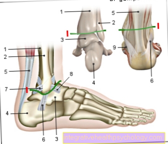
I - Upper ankle
(Joint line green) -
Articulatio talocruralis
- Shin -
Tibia - Fibula -
Fibula - Ankle bone -
Talus - Heel bone -
Calcaneus - Achilles tendon -
Tendo calcaneus - Fibula-calcaneus tape -
Calcaneofibular ligament - Hint. Shin-fibula
Ligament (rear syndesmosis ligament)
Posterior tibiofibular ligament - Front Fibula ankle ligament - Ligamentum fibulotalare anterius
- Delta band - Deltoid ligament
You can find an overview of all Dr-Gumpert images at: medical illustrations
Lower ankle illustration
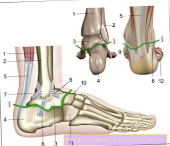
I - Lower ankle
(Joint line green) -
Articulatio talocalcaneonavicularis
- Shin - Tibia
- Fibula - Fibula
- Ankle bone - Talus
- Heel bone - Calcaneus
- Achilles tendon -
Tendo calcaneus - Fibula-calcaneus tape -
Calcaneofibular ligament - Hint. Shin-fibula
Tape-
Posterior tibiofibular ligament - Front Fibula ankle
Tape-
Lig fibulotalare anterius - Delta band - Deltoid ligament
- Scaphoid bone - Navicular bone
- Cuboid bone -Os cuboideum
- Fibula short muscle - Musculus fibularis brevis
You can find an overview of all Dr-Gumpert images at: medical illustrations
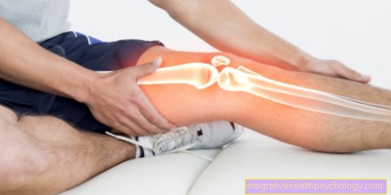
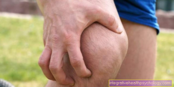
Chipped bone in the knee
A bone splintering in the knee area is mostly caused by direct Violencee.g. during sport or in traffic accidents. The patients mostly complain about strong pain and often over one limited joint mobility.
Chipped bone in the knee is usually treated operational, especially if accompanying other injuries, such as broken bones or Ligament injuries exist. On the other hand, the fragmented piece of bone is only very shifted slightly and do not lie Accompanying injuries before, can also be a non-operative therapy be considered.
In a way, one Special case of bone chipping on the knee represents the so-called Osteochondrosis dissecans It comes about, probably through a Reduced blood flowto slow down Death of a small area of bone under the articular cartilage.
In advanced stages this can develop Loosen the bone area and as a so-called "joint mouse" splinter into the joint capsule. In most cases, however, this complication can be avoided by consistently protecting the knee joint.
The most important distinguishing features from the other types of bone splintering in the knee are slowly progressive increase in pain intensity as well as arising without a sports or other accident as an acute cause of injury as well as conservative therapy in the form of rest.
Figure knee joint
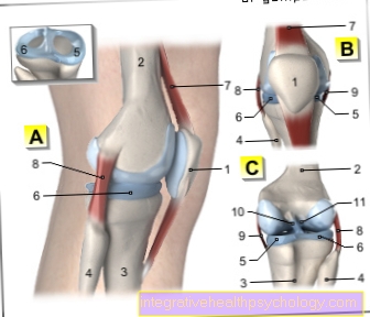
A - Right knee joint from the left
B - Right knee joint from the front
C - Right knee joint from behind
- Kneecap - patella
- Femur - Femur
- Shin - Tibia
- Fibula - Fibula
- Inner meniscus -
Meniscus medialis - Outer meniscus -
Lateral meniscus - Kneecap ligament -
Ligamentum patellae - Outer band -
Ligament collaterale fibulare - Inner band -
Ligament collateral tibial - Posterior cruciate ligament -
Ligament cruciatum posterius - Anterior cruciate ligament - Ligament cruciatum anterius
You can find an overview of all Dr-Gumpert images at: medical illustrations
Chipped bones on the fingers
A chipped bone in the area of the fingers is usually a sports injury. It often arises from a failed attempt to catch a heavier ball or from the ball hitting the tip of an outstretched finger. In addition to the fragmentation of bone, a dislocation of a finger joint can also occur.
Chipped bones in the finger can also be treated conservatively or surgically, depending on their severity. Since the fingers are very delicate parts of the body, great importance should be placed on the bone splinter to heal well again. If there is an accompanying injury to one of the finger tendons, this is of great importance. In particular, injuries to the flexor tendons must always be treated surgically, otherwise the ability of the finger to grasp can be permanent.
A peculiarity of a bone fragment on the finger is the constant stress on the fingers in everyday life. This makes it particularly difficult for the patient to adhere to the rest period required after the treatment. However, since insufficient rest can lead to long-lasting symptoms and thus jeopardize the success of the therapy, a high level of patient discipline is the key to success here.
Further information also under our suitable topics:
- Dislocation of a finger joint
and - Capsule tear on the finger
Chipped bone on the thumb
A chipped bone in the thumb mostly results from accidents in everyday life or sports. It occurs particularly frequently with goalkeepers in ball sports, with volleyball or basketball players or in the context of a so-called "ski thumb".
In the case of a “skier's thumb”, the outer collateral ligament in the base of the thumb tears due to the leverage of the thumb ligament of the ski pole, which leads to an unnaturally strong splaying of the thumb.
In more unfortunate cases, this torn ligament is associated with a bone fragment. In addition to unspecific signs such as pain and swelling, an increased ability to expand is typical for the skier's thumb, which is usually restricted by the now torn collateral ligament. In addition, patients find it difficult to hold objects between thumb and forefinger. Since the rupture of the collateral ligament alone - possibly after surgical treatment - is immobilized for several weeks, the bone fragment is also optimally treated in this way. Only in the case of a complicated, i.e. strongly displaced bone fragmentation, should surgical intervention be undertaken in order to ensure that the piece of bone grows back onto the remaining bone without complications.
A bone splintering in the thumb can, however, also develop independently, unrelated to a skier's thumb. It is based on similar injury mechanisms as the skier's thumb. In the vast majority of cases, surgical therapy can be dispensed with, and after four to six weeks of immobilization, the bone splinter should have healed to such an extent that the thumb can initially be subjected to everyday stress again, before the patient also engages after about 3-4 months can approach high-risk sports like skiing or basketball again in this regard.
The Rolando fracture can be viewed as a special form of bone splintering in the thumb. This mostly results from similar accidents as the injuries described above and refers to a fracture within the metacarpophalangeal joint of the thumb in combination with the splitting off of a piece of bone due to the tensile effect of a muscle tendon attached to it. Strictly speaking, the Rolando fracture is a metacarpal fracture, but due to the similar symptoms and injury mechanisms, it should be considered as a possible accompanying injury when diagnosing a bone splinter in the thumb.
For more information, see our topic:
- Ski thumb
and - Capsule tear on the thumb


.jpg)