Lymphatic vessels
Anatomy of the lymphatic vessels
The lymphatic vessels are anatomical structures that, like blood vessels, run through the entire body. Just like the bloodstream, the lymphatic vessels also carry a fluid.
As the name suggests, the lymphatic fluid is transported through the lymph vessels. The anatomy of the lymph vessels is very similar to the anatomy of the blood vessels, with the difference that there are always lymph nodes interposed between the individual lymph channels.

To understand the anatomy of the lymphatic vessels, one must first understand theirs function understand.
Transport the lymph vessels Tissue fluid (lymph) together with the included Proteins and white blood cells (Lymphocytes) from the periphery of the body towards the center. Roughly speaking, the periphery is everything that is further away from the heart (legs and arms, i.e. the extremities). From there the liquid gets over that Lymphatic vessels transports and flows into the area of the heart Venous angle (Confluence of the Internal jugular vein and the Subclavian vein to Brachiocephalic vein).
The anatomy of the lymph vessels is very similar to the anatomy of the veins, with one crucial difference. While the blood flow from arteries and veins is always connected and not interrupted, the lymphatic system has so-called blind ends on. This means that the lymph vessels start blindly with an open end in the tissue, much like a straw that is open on one side.
These lymphatic vessels, which begin blind in the periphery, become Lymph capillaries or initial lymphatic vessels called. These are extremely narrow vessels, which in the Intercellular spaces and can absorb the tissue fluid from there. The anatomy of the lymphatic vessels thus begins with a special feature. There are also capillaries in the blood system, but they are connected to one another. The lymph vessels, however, lie open in the fabric and can thereby absorb the fluid from the intercellular spaces.
Small anchor filaments are attached to the lymph vessels, which ensure that the vessel cannot slip. In addition, these filaments ensure that the interior (Lumens) the lymph vessels remain open and the fluid can flow in.
Precollateral
The anatomical structure of the lymph vessels following the lymph capillaries are the so-called Precollateral. These arise when several of the 50 µm wide lymph capillaries combine to form an approximately 100 µm wide lymph vessel. This thus represents one Confluence of several lymph capillaries and with the help of muscle cells it transports the fluid towards the left breast. In addition to the transport function, the precollaterals also serve to accommodate others Lymphliquid from the surrounding tissue. The anatomy of the lymphatic vessels is therefore quite simple.
Collaterals
Next, several precollaterals merge into one larger collector lymph vessel (or collateral lymph vessel). In comparison to the capillaries and the precollaterals, the collaterals serve exclusively to transport the lymph fluid onward. No more fluid is absorbed from the tissue.
These collaterals each have a diameter of 150 to 600 µm. The anatomy of these lymph vessels is almost identical to that of the veins. The collaterals have the histological classic three-layer wall structure (Intima, media and externa) and also have flaps, which ensure that the liquid is transported towards the left chest and does not sink into the arms or legs. The area between two valves is called the lymphatic vessels Lymphangion designated. This area contracts 10-12 times per minute and thus ensures that the lymph is transported further.
A total of 3 sub-forms of collaterals can be distinguished.
- The superficial (epifascial) The system is located in the subcutaneous fatty tissue and absorbs the lymph from the skin and fatty tissue.
- The deep (subfascial) The system found in the arms and legs (extremities) and in the trunk absorbs the lymph from muscles, ligaments, joints and bones.
- Finally, the viscera system follows (visceral system)which absorbs lymph from the various organs.
The anatomy of the lymph vessels also ensures a connection between these three systems. Thus, the lymph can flow from the deep system into the superficial system. The connection between the vessels is called Anastomosis or Perforation cycle designated.
Lymphatic collection points
A specialty of the anatomy of the lymph vessels are the Lymphatic collection points. These are the largest lymph vessels in the human body. They are divided into the upper or lower half of the body depending on the position.
They include the Tracheal trunk (Truncus trachealis), as well as the Milk breast duct (Ductus thoracicus), which is about 40 cm long. These collection points take in lymph from the collaterals. Then they flow into the in the area of the heart left venous angle a. At this point, the anatomy of the lymphatic vessels connects with the anatomy of the venous system.
Lymph vessel valves
The structure of the lymph vessels is generally very similar to that of the Veins, especially with the larger lymph vessels (Collaterals). Similar to the veins, the lymphatic vessels also have one three-layer wall structure, which classically one Intima, a media and an external consists.
Another similarity is that Valves of the lymphatic vessels. As with the veins, the valves of the lymph vessels ensure that the liquid (Lymph) can be transported from the periphery, for example from the leg, towards the left chest. Since the fluid has to flow in the opposite direction to gravity, the lymph vessels need valves to ensure adequate flow and a To prevent reverse current. These flaps are located only in larger lymph vessels like that Collaterals, but not in the Capillaries and Precollateral. In contrast to the venous system, the valves are the lymphatic vessels passive. They are present in the larger lymph vessels at a certain distance and depending on their diameter.
It comes to one reduced function of the valves of the lymphatic vessels, it may be that the fluid can no longer be adequately transported and it leads to the formation of so-called Lymphedema comes. In general, a malfunction of the valves of the lymphatic vessels is compared to a decreased one Venous valve function rather seldom.
Figure lymphatic system

Lymphatic system
- Head lymph nodes -
Nodi lymphoidei capitis - Cervical lymph nodes -
Nodi lymphoidei cervicales - Mouth of the breast duct
in the left arm-head vein -
Thoracic duct
Left brachiocephalic vein - Mouth of the right main
lymph duct in the right
Arm head vein -
Dexter lymphatic duct
Vena brachiocephalica dextra - Superior vena cava -
Superior vena cava - Axillary lymph nodes -
Nodi lymphoidei axillares - Milk breast duct -
Thoracic duct - Lymphatic vessels -
Vasa lymphatica - Abdominal lymph nodes -
Nodi lymphoidei abdominis - Pelvic lymph nodes -
Nodi lymphoidei pelvis - Inguinal lymph nodes -
Nodi lymphoidei inguinales - Mandibular lymph nodes -
Nodi lymphoidei submandibulares - Anterior cervical lymph nodes -
Nodi lymphoidei cervicales anteriores - Superficial lateral cervical lymph nodes -
Nodi lymphoidei cervicales
lateral superficiales - Deep lateral cervical lymph nodes -
Nodi lymphoidei cervicales
lateral profundi - Mastoid lymph nodes -
Nodi lymphoidei mastoidei - Occipital lymph nodes -
Nodi lymphoidei occipitales - Facial lymph nodes -
Nodi lymphoidei faciales - Parotid lymph nodes -
Nodi lymphoidei parotidei
You can find an overview of all images from Dr-Gumpert under: medical images
Lymph vessels of the head
Transport the lymph vessels on the head Tissue fluid, Proteins and immune cells from the head to the left vein corner.
The tissue fluid then returns to the blood here.
Since the flow of lymph fluid in the head is directed downwards due to gravity and automatically runs back into the left vein angle, it comes here very rarely to lymphedema. On the way to left venous angle several pass through the lymph vessels of the head Lymph node regions on the neck.
If there is an infection in the head area, such as an ear infection or sinus infection, these cervical lymph nodes can swell. The reason for this is that the lymph fluid contained in the lymph vessels of the head is cleaned in the lymph nodes.
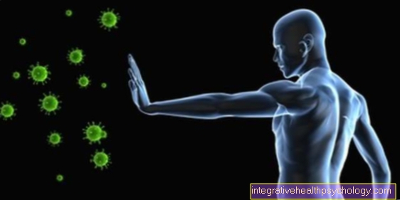
All inflammatory cells thus collect in the area of the cervical lymph nodes, so that it becomes active and increasingly produces immune cells against the pathogens (T lymphocytes and B lymphocytes).
It comes to one severe swelling of a cervical lymph node, it may be that the lymphatic fluid cannot drain off sufficiently and backs up in the lymphatic vessels of the head.
If so, the patient suffers from pale and sallow facial skin. There is rarely any redness or pain. Here one can Lymphatic drainage effectively help to ensure the outflow of lymphatic fluid again.
Lymph vessels of the brain
For a long time, researchers were not sure whether there were lymph vessels in the brain or whether the brain was free of any lymphatic vessels. It wasn't until about a year ago that researchers published a report that suggested that lymph vessels exist in the brain. You are in the outermost of the three meninges, the so-called Dura mater.
Their function has not yet been clarified with certainty, but it is assumed that these lymph vessels Immune cells in the direction of the brain and thus play a decisive role in defense against pathogens.
Furthermore, the brain's lymph vessels seem to play an important role when it comes to the so-called Cerebrospinal fluid (Brain water or cerebrospinal fluid) from the Cerebral ventricles to transport.
So far, lymphatic vessels in the brain have only been detected in mice. However, it is assumed that people in the meninges (Dura mater) Have lymphatics, which play a decisive role in the defense and possibly a new explanation for Disease Alzheimer because harmful metabolic products could also be transported into the brain via these pathways.
Lymph vessels of the face
Most of the time, lymphatic vessels are connected to the legs, as lymphedema can develop particularly quickly here. The actual function of the lymph vessels, namely the removal of fluid, is then no longer guaranteed.
But there are also lymph vessels in the face. Their job is to remove tissue fluid from the face and reinsert it into the venous system. Furthermore, they should transport proteins and immune cells such as lymphocytes. The lymph vessels of the face also ensure that pollutants are transported away.
If this function is disturbed, this can easily become noticeable. The patient looks pale, has a pale complexion, or has blemished facial skin. If there is a congestion of the lymph vessels in the face, this can be treated with the help of lymph drainage. This has the effect that the harmful substances are better transported away with the lymph fluid, which in turn can improve the appearance of the skin.
Read more on the subject here: Lymphedema.
Lymph vessels of the neck
With the help of lymph capillaries, the fluid is absorbed from the tissue on the neck (e.g. from the muscles) and transported away in the direction of the left or right venous angle in the area under the clavicle. This is where the fluid, which contains proteins and immune cells, flows into the venous system.
In the lymph node, the lymph fluid is cleaned of all harmful substances, which is why it can swell in the event of an infection. After this purification of the lymph fluid, it returns to the venous system via the lymph vessels in the neck and thus closes the circulation.
The lymph vessels in the neck have a particularly large number of lymph nodes. These lymph nodes become active when there is inflammation or infection, such as tonsillitis or the flu. Especially with the almonds (Tonsils), swollen lymph nodes on the neck are particularly visible from the outside.
Read more on the subject at: Lymph nodes on the neck.
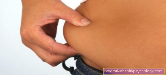
Breast lymph vessels
The lymph vessels of the breast have a special meaning. Just like on the neck, there are also in the chest area multiple lymph nodesin which the Lymph fluid can be cleaned. These play a major role Breast cancer (Breast cancer) plays a crucial role, which is why the breast lymph vessels are particularly important.
In general, the lymph vessels of the right breast in the right venous angle. The lymph vessels of the left breast however flow into the left venous angle. Before the fluid is transferred into the venous system, the lymph vessels pass through the Lymph nodes in the armpit area as well as under the Collarbone.
At a Breast cancer the tumor cells are taken up by the lymph vessels. This then gets them to the lymph nodes, especially in the armpit area. As a result, the tumor is not only located in the breast, but also in the lymph node. Therefore, during an operation every lymph node removed infected by tumor cells.This peculiarity of the breast lymph vessels is of crucial importance in breast cancer. The Number of affected lymph nodes is crucial for the prognosis of the tumor.
- Lymph node swelling in the armpit - dangerous?
- Breast cancer signs
Lymph vessels of the arm and hand
Lymph vessels are also present in the arm and hand. These have the typical structure as in the rest of the body and transport the tissue fluid from the skin, fat, muscles and bones of the arm and hand.
A special feature is that only the lymph fluid from the right arm and the right breast enters the right venous angle merge. The lymph fluid from all other parts of the body (for example the right leg) flows into the areas near the heart left venous angle.
The lymph vessels of the arms and hand pass through two larger ones Lymph node stations. These are, on the one hand, the lymph node station in the elbow area and the lymph node station in the armpit area. In the event of an injury, for example to the hand, it can lead to swelling these lymph node stations come (see: Lymph nodes swollen - how dangerous is it?)
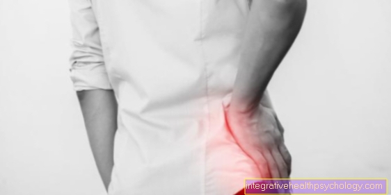
Lymph vessels of the leg

The lymph vessels have the task of transporting fluid, for example in the leg, towards the left breast and transferring this fluid to a vein near the heart (Brachiocephalic vein) to hand over.
The lymph vessels of the legs are in this respect especiallybecause they are furthest from this vein. Since, due to the force of gravity, the lymph fluid would normally flow from the upper body towards the legs, the lymph vessels must flow towards the legs with the help of Muscles and valves make sure this Backflow prevented becomes.
However, it comes to one increased pressure in the veins, for example due to a weak heart (Heart failure), this pressure may be transferred to the lymphatic system. Since the lymph vessels of the legs have to fight particularly hard against gravity, additional pressure has a negative effect on the vessels. You can no longer adequately pump the fluid from the leg towards the heart, which then causes it to become so-called in the legs Lymphedema can come. This lymphedema always occurs when the lymph vessels of the legs are overloaded and can no longer prevent the fluid from flowing back into the leg.
Inflammation of the lymph vessels
An inflammation of the lymph vessels (also Lymphangitis) is mostly caused by pathogens (bacteria) or other poisons (snake venom, insect venom, chemotherapeutic agents). When pathogens or pollutants that circulate in the blood get into the lymphatic system, this often leads to inflammation of the lymph vessels or the lymph nodes. Lymphangitis often develops on the basis of an infection Staphylococci or Streptococci.
If the lymphatic vessels are inflamed, they can be enlarged and palpable, or even visible (reddish stripes starting from the entry wound). In the process, the lymph vessels are also overheated and can be painful. In general, inflammation of the lymph vessels leads to a fever with chills and a feeling of weakness. In some cases, the heart may beat faster because of the infection (tachycardia). Lymph vessel inflammation can also spread in the lymph system and affect lymph nodes.
In the case of mild courses, immobilization and cooling of the affected parts of the body reduce the pain. Dressings and ointments can also improve the symptoms. In severe cases, the use of antibiotics to support the immune system is recommended.
Read more about this topic here: How dangerous is lymphangitis?


.jpg)