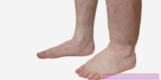
Metatarsus
General
The metatarsus consists of five metatarsal bones (Os metatarsalia I - V), which are connected to one another by joints. They are located in the foot between the toes and the tarsus. Each metatarsal bone forms a ray with the respective toes, dividing the entire foot into five rays. The first ray is formed from the big toe and the metatarsal bone that belongs to it, the fifth ray from the little toe and the metatarsal bone.
Figure ankle

- Toe phalanx -
Phalanx distalis - Middle toe -
Phalanx media - Phalanx -
Phal. proximalis
(1st - 3rd toe bones -
Phalanges) - Metatarsal bones -
Os metatarsi - Inner sphenoid bone -
Medial cuneiform bone - Middle sphenoid bone -
Os cuneiform intermedium - External sphenoid bone -
Os cuneiform laterale - Cuboid bone - Os cuboideum
- Scaphoid bone - Navicular bone
- Ankle bone - Talus
- Ankle roll - Trochlea tali
- Heel bone - Calcaneus
- Protrusion on the 5th metatarsal -
Tuberositas ossis metatarsalis quinti (V)
You can find an overview of all Dr-Gumpert images at: medical illustrations
construction
The metatarsus consists primarily of the five metatarsal bones, which all have the same basic structure.
Each metatarsal bone consists of a base, a body (Corpus) and a head (Caput, metatarsal head).
In the area of the base of the first metatarsal there is a bony process. This is called Metatarsal tuberosity I designated. This is where the tibialis anterior muscle attaches, which can lift the foot and turn it inward.
There is also an extension in the area of the fifth metatarsal Metatarsal tuberosity V.. This serves as an attachment for the peroneus brevis muscle, which can bend the foot and turn it outwards.
Both the bases and the heads of the individual bones have articular surfaces, which allows the metatarsal bones to interact with the adjacent bony structures.
The heads form a convex articular surface and interact with the adjacent toe base. They thereby form the five metatarsophalangeal joints.
The bases of the metatarsal bones are rather flat and are in contact with the tarsus.
The bases of the first through third metatarsals articulate with the three Ossa cuneiformia (Cuneiform bones), the bases of the fourth and fifth metatarsals with the cuboid bone.
The is formed between the tarsus and the bases of the metatarsals Lisfranc joint line out.
In addition, the individual bases of the metatarsals have contact with each other and thus form the Intermetatarsal joints.
In the area of Metatarsophalangeal joint lie two Sesame bones. These are embedded in the tendons and the capsular ligament apparatus.
On the middle sesamoid, the Abductor hallucis muscle and part of the Flexor hallucis brevis muscle on. Both muscles are used to flex the big toe and maintain the longitudinal arch of the foot, while the abductor is used to spread the big toe.
The Adductor hallucis muscle on. This keeps the transverse arch of the foot upright and can place the big toe on the foot. In addition, the second part of the Flexor hallucis brevis muscle on this sesamoid bone and bends the big toe in the base joint.
Appointment with ?

I would be happy to advise you!
Who am I?
My name is I am a specialist in orthopedics and the founder of .
Various television programs and print media report regularly about my work. On HR television you can see me every 6 weeks live on "Hallo Hessen".
But now enough is indicated ;-)
Athletes (joggers, soccer players, etc.) are particularly often affected by diseases of the foot. In some cases, the cause of the foot discomfort cannot be identified at first.
Therefore, the treatment of the foot (e.g. Achilles tendonitis, heel spurs, etc.) requires a lot of experience.
I focus on a wide variety of foot diseases.
The aim of every treatment is treatment without surgery with a complete recovery of performance.
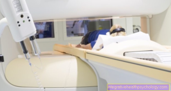
Which therapy achieves the best results in the long term can only be determined after looking at all of the information (Examination, X-ray, ultrasound, MRI, etc.) be assessed.
You can find me in:
- - your orthopedic surgeon
14
Directly to the online appointment arrangement
Unfortunately, it is currently only possible to make an appointment with private health insurers. I hope for your understanding!
Further information about myself can be found at
function
The metatarsus is part of the whole Foot at the Construction and maintenance of the longitudinal and transverse vaults involved. In addition, the individual metatarsals can be moved relative to each other and thus allow the Inward and outward rotation of the footso that it can adapt to bumps while walking. In addition, the metatarsals are Starting point of various muscles whereby the flexion and extension of the entire foot and the movements of the big toe are possible.
Diseases
in case of an hallux valgus or one splayfoot there are changes in the position of the metatarsals.
The metatarsal bones can break when direct force hits the foot from the front.
In addition to this pattern of injury, the metatarsal bones can also break under constant stress on the metatarsus. This type of break is called a Fatigue fracture designated.
You can find further information under our topic: Metatarsal fatigue fracture
Summary
The metatarsus consists of five bones, Metatarsal bone IVwho have favourited various articulated connections to have the adjacent bones of the foot. This ensures the mobility of the foot and the adaptation to uneven ground when walking. In addition, various Muscles to the bone of the metatarsus, which ensure both mobility and tension and posture of the longitudinal and transverse arches.
The very frequent misalignment of the big toe (hallux valgus) is caused by a misalignment of the metatarsal bones, just like the splayfoot. Also Fractions are possible in the metatarsus that can lead to a fracture through direct force or constant stress.