Nasal cavity
introduction
The nasal cavities are counted among the upper air-conducting airways. It is made up of bony and cartilaginous structures. In addition to the respiratory function, it is relevant for antibacterial defense, language formation and olfactory function. It is associated with various structures in the skull area.
Illustration of the sinus and nose

- Frontal sinus -
Frontal sinus - Ethmoid cells -
Cellulae ethmoidales - Maxillary sinus -
Maxillary sinus - Sphenoid sinus -
Sphenoid sinus - Thin septum -
Septum sinuum frontalium
You can find an overview of all Dr-Gumpert images at: medical illustrations

- Upper turbinate -
Concha nasi superior - Upper nasal passage -
Superior nasal meatus - Middle turbinate -
Concha nasi media - Middle nasal passage -
Meatus nasi medius - Lower turbinate -
Concha nasi inferior - Lower nasal passage -
Inferior nasal meatus - Atrium of the nasal cavity -
Vestibulum nasi - Olfactory threads - Fila olfactoria
- Olfactory bulb - Olfactory bulb
- Rear opening of the
Nasal cavity - Choana - Nasal cavity - Cavitas nasi
- Pharyngeal almond -
Pharyngeal tonsil - Frontal sinus - Frontal sinus
- Sphenoid sinus -
Sphenoid sinus - Oral cavity - Cavitas oris
- Tongue - Lingua
You can find an overview of all Dr-Gumpert images at: medical illustrations
Anatomy of the nasal cavity
The nasal cavity opens after ventral (front) over the two nostrils (Nares). She goes over the back Choanae in the Pharynx (throat) above. To lateral (to the side), after cranial (above) and after caudal (below) it is limited to the bones.
The front part of the nasal cavity is called Verstibulum nasi (Nasal vestibule). It extends from the nostrils (nares) to an arched fold of the mucous membrane, which forms the transition to the main nasal cavity (Cavitas nasi propria) represents. At this fold of the mucous membrane (Limen nasi) is the narrowest section (in the outer nasal part) of the anterior nasal cavity.
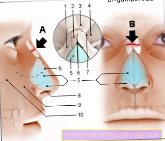
The typical outer shape of the nose is made up of the nasal cartilage and the bony bridge of the nose (Radix nasi) educated. The nasal cartilage forms the Bridge of the nose, the Nasal septum (Nasal septum) and the Nostrils. It consists of several cartilaginous parts. The Cartilago alaris major (large alar cartilage) forms the border of the nostrils and the nostrils with the Crus mediale (between the nostrils / nasal bridge) and Lateral crus (outside around the nostrils) - including the tip of the nose. The nostrils are additionally still through the Cartilagines alares minores (small alar cartilages) formed. The Cartilago septi nasi forms the cartilaginous septum nasi (nasal septum), which the Nasal vestibule divides into two parts (one on the right and one on the left). From the Limen nasi then begins the Nasal cavity.
This is after lateral (to the sides) through the Conchae nasales (Nasal concha) limited. The Conchae nasales are bone protuberances (bone lamellae) of different skull bones: parts of the Ethmoid bone (Ethmoid bone), of Upper jaw (Maxillary bone), of Palatine bone (Palatine bone) and des Tearbone (Lacrimal bone). Between the conchae nasales lie on the lateral nasal wall Nasal passages. They open dorsally over the two choans (funnels) into the throat. The nasal passages themselves represent an opening area for ducts and sinuses.
There are three nasal passages (meatus nasi):
- The uppermost nasal passage (Superior nasal meatus) connects the nose with the Sphenoid sinus (Spenoidal sinus); this is one of the Sinuses. He also provides the muzzle for the posterior ethmoid cells These are air-filled bone cavities (Pneumatization rooms) in the skull. In addition, the human olfactory organ is located in the superior nasal meatus.
- The middle nasal passage lies on the side and under the medial nasal concha. The other paranasal sinuses open over it (Frontal and maxillary sinuses), as well as like that anterior and posterior ethmoid cells.
- The lower nasal passage (meatus nasi inferior) provides the connection to the Lacrimal system the lacrimal duct (Durctus nasolacrimalis) ends here.
If that eye tears, she runs Tear fluid through a system of Tearways finally into the lacrimal duct and via the meatus nasi inferioir into the nose. If there is little tear production, the liquid evaporates after it emerges in the nose. With increased tear production, e.g. when people cry a lot, it happens that they have the feeling that they are swallowing their tears. This is because the inferior nasal meatus is close to the choanas, so that the tear fluid flows through them into the Nasopharynx expires and thus in the neck.
Up is the Nasal cavity by the Nasal roof limited. This is formed from parts of the sphenoid bone, des Nasal bone, ethmoid, and Frontal bone. Here the nasal cavity stands above that Sphenopalatine foramen (a bone opening) in connection with the wing bone fossa. This is a bony depression between two protrusions on the upper jaw (Maxillary bone) and cuneiform (Sphenoid bone). Pull through this foramen annoy and vessels that are responsible for supplying the nasal cavity.
The nasal cavity is delimited at the bottom by parts of the upper jaw, the intermaxillary bone and the palatine bone. Here is the one Canalis incisivus - a bony canal that connects the nasal cavity with the Oral cavity connects. Nerves and vessels pass through it to supply the palate. The middle nasal wall, the nasal septum (Nasal septum), divides the nasal cavity into a left and right section. In the anterior part, the nasal septum is formed in a cartilaginous manner. In the back, the nasal septum is bony. If the nasal wall is unevenly positioned, one side of the nasal cavity can be so narrow that the flow of air is obstructed. Here one can operative treatment be necessary.
histology
The nasal cavity can histologically (microscopically) divided into three parts:
- The Skin region lies in the nasal vestibule and resembles the skinthat lies on the outside of the nose. She is with Hair and many sweat and sebum glands occupied. There are also large ones here Veins in the nasal wall.
- The Nasal cavity consists of two different ones Mucosal types.
- On the one hand, the respiratory epithelium; this is the characteristic one multi-row high prismatic epithelium of the respiratory tract, which is covered with goblet cells and cilia (Kinocilia) is busy. Kinocilia are cell protuberances that are mobile and thus ensure that foreign bodies and dirt are transported away (towards the throat). Goblet cells support the removal through the production of a thin (serous) Mucus. In addition, the air you breathe is moistened by the formation of slime.
- On the other hand, there is the in the main nasal cavity Regio olfactoria. It only makes up a portion of the entire nasal cavity mucosa the size of a thumbnail. It lies on the roof of the nose and on the upper turbinate. The olfactory region is part of the imperial organ - it represents the surface through which odorous substances are registered and the specific information to the brain is forwarded. To do this, it has special olfactory cells, which are counted among the sensory cells, and have binding sites for odorous substances on their surface.
Function of the nasal cavity

One of the main tasks is to direct the air you breathe. This is warmed and moistened in the nasal cavities. The warming takes place via a distinctive network of vessels in the nose, in which warm blood flows, which gives off part of the warmth to the inhaled air.
The air in the nasal vestibule can be cleaned of contaminants through the hair in the nasal vestibule, which can be removed from the goblet cells by the movements of the cilia and the mucus.
In addition to catching dirt, one of the tasks is to render bacterial pathogens harmless. This is possible because the mucus produced contains antibacterial components and immune cells are also located in the mucous membrane.
Furthermore, part of the speech production takes place via the nasal cavity together with the paranasal sinuses. The cavities in the skull function as resonance spaces.
In addition, the human olfactory perception of smell runs through the olfactory region. In this context the Jacobson organ (Vomeronasal organ) - this is only rudimentary in humans. These are also olfactory cells. However, these are responsible for the perception of pheromones (fragrances that unconsciously influence sexual behavior).
Read on under: Pheromones for men




.jpg)
.jpg)








.jpg)

