Osteosarcoma
All information given here is only of a general nature, tumor therapy always belongs in the hands of an experienced oncologist!
Synonyms
Bone sarcoma, osteogenic sarcoma
English: Osteosarcoma
definition
The osteosarcoma is a malignant bone tumor that belongs to the group of primarily osteogenic (= bone-forming) malignant (= malignant) tumors.
According to statistical surveys, osteosarcoma is the most common malignant bone tumor. It was also possible to determine an increase in the occurrence in growing age, although adults can also contract the disease.
Osteosarcomas tend to metastasize early on.
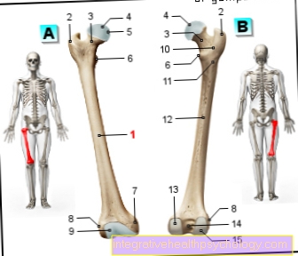
With regard to the localization of an osteosarcoma, it was found that growth plates of long tubular bones, such as the ulna and radius, are typically affected. An involvement of the spine of the knee (= 50% of all osteosarcomas) and hip joint etc. are also conceivable.
In the course of tissue examinations (= histological examinations) it was found that osteosarcomas consist of so-called polymorphic bone-forming cells and.
Summary

As mentioned above, osteosarcomas are malignant tumors:
There are different subgroups of an osteosarcoma. Depending on the location or origin:
- the osteogenic sarcoma, which starts from the bone.
- Osteosarcomas with a tendency to ossify or to form osteoid tissue (= osteoid sacroma)
In the course of histological examinations it was found that in the case of an osteosarcoma disease there are bone cells that can no longer produce the basic bone substance (bone calcification). Such so-called tumor cells have the property of spreading. They don't respect cell boundaries.
As already mentioned in the context of the definition, osteosarcomas tend to occur in the growth gap. About 50% of all diagnosed osteosarcomas are found in the knee joint. Other locations can be: ulna, radius, hip joint, Spine, ....
Osteosarcomas are prone to metastasis. The formation of metastases (= colonization of other areas of the body with tumor cells) is particularly common in the area of the lung, or into the lymph nodes. The colonization of the lymph nodes is much less common. If the disease is discovered early enough, metastasis can be avoided.
The symptoms in the early stages of the disease are initially not indicative, however, due to the radical growth behavior of an osteosarcoma, symptoms such as (strong) Pain and Swelling a. These symptoms have to be differentiated from a differential diagnosis. Often there is initially the suspicion that the bone is inflamed (Osteomyelitis).
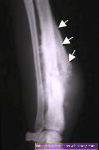
X-ray examinations can be performed to establish the diagnosis. In addition, any metastases can be eliminated through a 3 - phase Scintigraphy prove. This diagnostic method is used in particular to check the success of chemotherapy or for follow-up checks (to rule out recurrences). Often that happens too CT for use. A CT can be used to estimate the extent of the tumor. In particular after chemotherapy, angiography (= X-ray diagnostic representation of the (blood) vessels after injection of an X-ray contrast agent) can also be performed. In order to determine whether the tumor is malignant or not, tissue is taken and examined as part of a biopsy.
The therapy is usually divided into two phases:
- chemotherapeutic pretreatment
- surgical removal of the tumor
This two-part therapy significantly increases a patient's prognosis. With only surgical therapy, the probability of healing was (only) 20%. In the corresponding section, the form of therapy will be discussed in more detail.
It is currently unclear which factors favor the occurrence of osteosarcoma. As with almost all other bone tumors, hormonal and growth-related factors are suspected to be triggering factors.
An osteosarcoma develops from one rather rarely M. Paget, or after radiotherapy or chemotherapy for another existing disease. According to statistical surveys, however, an increased likelihood of osteosarcoma after illness with retinoblastoma (tumor in eye in children).
The forecast cannot be formulated across the board. A prognosis for osteosarcoma is always dependent on many individual factors, such as time of diagnosis, initial tumor size, location, metastasis, response to chemotherapy, extent of tumor removal, etc.
However, it can be said that due to the changed form of therapy (see above) a Five year survival rate of about 60% can be achieved.
Occurrence
The peak of the disease is during puberty, which means that osteosarcomas occur very frequently in children and adolescents, usually between the ages of 10 and 20 years.
Mostly male adolescents are affected by the disease.
Osteosarcomas represent around 15% of all primary malignant bone tumors, so osteosarcoma is the most common malignant bone tumor in (male) children and adolescents.
Osteosarcomas can also develop in adults. This is usually the case if previous illnesses such as Paget's disease (=Osteodystrophia deformans Paget) occurred. It is also possible that the clinical picture develops after chemotherapy or radiation therapy.
causes
As already mentioned in the summary, the causes for the development of an osteosarcoma have not yet been adequately clarified.
As with almost all other bone tumors, hormonal and growth-related factors are suspected to be triggering factors.
An osteosarcoma develops from one rather rarely M. Paget, or after radiotherapy or chemotherapy for another existing disease. According to statistical surveys, however, there was an increased likelihood of an osteosarcoma after illness Retinoblastoma (Tumor in the eye in children)
metastasis
Due to the slope of the Osteosarcoma too early metastasis, an early diagnosis is of elementary importance. Metastasis is usually hematogenous, i.e. via the bloodstream. Above average, metastases are mainly found in the area of the lung, but also in the skeletal area (extension to other bones) or the Lymph nodes.
Since an early diagnosis is seldom made due to the poorly indicative symptoms, metastases are very often found as soon as the diagnosis is made. Statistically, this is the case for about 20% of all osteosarcoma patients.
It is assumed that micrometastases could already be detected in far more patients at the time of diagnosis. However, they are still too small so that they cannot be detected / displayed with the currently common diagnostic methods.
These micrometastases are attempted as part of the two-part form of therapy (see: Therapy)
- chemotherapeutic pretreatment
- surgical removal of the tumor
by means of chemotherapy kill.
diagnosis
The symptoms are often not indicative in the early stages. It kick first
- Pain and
- local signs of inflammation (redness, swelling, overheating)
on. In the further course, general symptoms of a tumor, such as:
- Swelling of the lymph nodes
- unwanted weight loss (more than 10% in 6 months)
- Paralysis
- Fracture without an accident event (pathological fracture)
- Night sweats
- paleness
- Loss of performance
to be added.
The diagnostic possibilities extend to
X-ray diagnosis:
Here is a X-ray examination made in the symptomatically conspicuous area (at least 2 levels).
Sonography:
The Sonography is particularly used when an osteosarcoma has already been diagnosed. It is used for differential diagnostics, in particular to delimit a soft tissue tumor.
general laboratory diagnostics (blood test):
- Blood count
- Determination of ESR (= sedimentation rate)
- CRP (C-reactive protein)
- Electrolytes
- Alkaline phosphatase (aP) and bone-specific aP:
- Prostate-specific antigen (PSA) and acid phosphatase (sP). These values are increased in prostate cancer, which in turn often metastasizes to bone.
- Iron: The iron levels are typically low in tumor patients
- Total protein
- Protein electrophoresis
- Urine status: paraproteins - evidence of myeloma (plasmacytoma)
Special tumor diagnostics:
Magnetic resonance imaging (MRI):
The Magnetic resonance imaging (MRI) can be used in addition to the imaging methods mentioned in the context of basic diagnostics.
Since an MRI shows the soft tissue particularly well, if an osteosarcoma is diagnosed, it is possible to assess the extent of the tumor to neighboring structures (nerves, vessels) of the affected bones, and thus also to estimate the tumor volume and clarify the local extent of the tumor.
If a malignant bone tumor is suspected, the entire diseased bone should also be imaged; if necessary, further diagnostic measures should be taken to rule out metastasis to other areas (see above).
Computed Tomography (CT):
A CT can be used to estimate the extent of the tumor.
Digital subtraction angiography (DSA) or angiography:
Angiography is the X-ray diagnostic representation of the (blood) vessels after injection of an X-ray contrast medium. In digital subtraction angiography, vessels (arteries, veins and lymphatic vessels) are examined using X-ray diagnostics.
Read more about the topic here Angiography
Skeletal scintigraphy (3-phase scintigraphy):
This is understood to mean an imaging procedure using radionuclides that are as short-lived as possible (e.g. gamma rays) or so-called radiopharmaceuticals. The skeletal scintigraphy is used to examine bones with regard to zones with increased bone metabolic activity or blood flow. They can provide information about the presence of osteosarcomas.
Biopsy:
To differentiate whether the tumor is malignant or not, tissue is removed and examined as part of a biopsy (= histopathological (= tissue) examination. A biopsy is often done if a tumor is suspected or if the type and severity of a tumor are unclear. Such an investigation could, for example, by means of Incisional biopsy can be performed. In doing so, the tumor becomes partial surgically exposed and a tissue sample (usually bone and soft tissue) removed. If a quick section analysis is possible, the removed tumor tissue can be examined and assessed directly for dignity.
Read more about the topics here: Osteosarcoma therapy and biopsy
forecast
The forecast cannot be formulated across the board. A prognosis for osteosarcoma is always dependent on many individual factors, such as the time of diagnosis, initial tumor size, location, metastasis, response to chemotherapy, and the extent to which the tumor was removed.
However, it can be said that the modified form of therapy (see above) can achieve a five-year survival rate of around 60%.
Aftercare
As recurrences cannot be ruled out, follow-up care should be provided. The following Follow-up recommendation can be pronounced, in individual cases a follow-up plan can also deviate from this:
- 1st and 2nd year
A clinical examination should be carried out every quarter. This clinical examination usually includes a local X-ray check and laboratory tests. Furthermore, a CT of the chest and a whole-body skeletal scintigraphy will be made. An MRI is usually performed every six months for the first two years.
- 3rd to 5th year
The clinical examination is now carried out every six months. Likewise, within the first and second year after the illness, the clinical examination usually includes a local X-ray check and laboratory tests. Furthermore, a CT of the chest and a whole-body skeletal scintigraphy will be made. A local MRI is now performed annually.
- from the 6th year
The clinical examination is usually carried out once a year. It includes a local X-ray control and laboratory tests. Furthermore, a CT of the thorax (chest) as well as a whole-body skeletal scintigraphy and a local MRI are made.
Further information
Further information on this topic can be found at:
- Osteosarcoma Therapy
- Bone cancer
There are different forms of bone cancer.
You can find further information on the following bone tumors:
- Osteoid osteoma
- Osteochondroma
- Chondrosarcoma
- Enchondroma
- Rhabdomyosarcoma
General information on the subject of tumors can be found at:
- Bone tumor
- tumor
All topics that have been published on the field of internal medicine can be found at:
- Internal medicine A-Z


