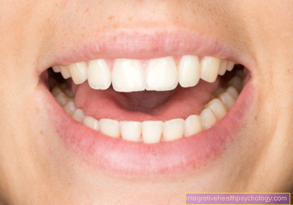
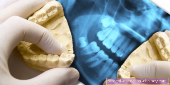
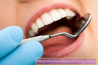
X-ray of teeth
introduction
The roentgen (or X-ray diagnostics) provides a possibility to radiate through the body and under the skin make visible structures.
The resulting images of this dental diagnostics can on special X-ray films or recorded as digital on the computer and then evaluated.
The radiation dose and the type of exposure must be specifically adapted for almost every part of the body, which means that the radiation exposure is different for different X-rays. Whereby the Radiation exposure from X-rays in dentistry in general and especially in comparison to CT vanishingly small is. See matching: Radiation exposure in CT
Also in the Dentistry the X-ray serves as an enormous aid in the Diagnosis, carious defects, inflammation, fractures and other anomalies of the bony Jaws can be represented exactly.
Standard procedures in dentistry include Dental films (also E.inzelzahnacalled recording), Bitewing recordings and the so-called OrthopantomoGramm (short OPG or OPT).
Panoramic image or OPG
The Orthopantomogram is often also called Panoramic layer recording and is a radiographic overview of the upper and lower jaw.
All teeth, all jaw sections, both Temporomandibular joints and the adjacent maxillary sinuses are shown in a single large x-ray image.
Used for recording at Ortopantomogram a X-ray equipment into which a film cassette or a digital line scan camera can be inserted.
During the delivery of the radiation, the receiving unit moves in a semicircle around the patient's head and thereby generates a Panoramic shot of the jaw.
This is done in a similar way to creating a panorama image with a normal camera, only that X-rays are used here and structures below the skin can be made visible. The radiation exposure for the patient is relatively low with this X-ray procedure.
Bitewing recording
Bitewing recordings are mainly used to find Caries and tooth decay not visible from the tooth surface. In addition, they provide an ideal overview of the situation of the tooth support system and help the therapeutic process of a so-called Periodontal treatment to plan. The radiation exposure is also relatively low with this type of X-ray.
Either Ortopanthomograms, as well as Bitewing recordings offer a good overview of the overall situation of the jaw, but are comparatively fuzzy and therefore not necessarily accurate in detail.
Tooth film or EZA
Images of individual teeth are called Dental films. When making such a single image, a so-called tooth film is placed directly behind the teeth and a freely rotatable X-ray source is placed outside the mouth so that the desired area is ideally displayed.
In contrast to the ortopanthomogram or the bitewing recordings, dental films impress with their unsurpassed quality Level of detailThe smallest defects can be shown particularly sharply and thus ideally assessed.
The roentgen Often several dental films are made of the teeth in order to be able to assess the entire jaw, similar to an ortopanthomogram.
This method is no longer used due to the comparatively high radiation exposure in X-rays. Dental films are only made to assess individual teeth or groups of teeth, only to gain an overview OPGs or bitewing recordings recorded. Only in the context of a periodontal disease are several EZAs still used today.
Can you X-ray your teeth during pregnancy?
It is also possible to X-ray the pregnant woman's teeth during pregnancy. The x-ray exposure that occurs when x-raying a tooth is between 0.003 and 0.054 mSv, depending on the imaging technique. The following applies: the more modern the X-ray machine, the lower the radiation exposure. Theoretically, damage to the unborn child is only to be expected from 30 mSV, which means that a pregnant woman's tooth would have to be x-rayed more than 500 times before the fetus could be damaged.
Learn more at: X-ray during pregnancy
However, since fetal damage can never be ruled out, it is advisable to only take X-rays during pregnancy in an emergency. This is the case, for example, if the mother is in mortal danger or if the toothache is very severe. If the intake cannot be bypassed, the exposure should be kept as low as possible. For this, the lead apron must be put on correctly and the recording technique used with the lowest possible dose.
You might also be interested in this: Toothache in pregnancy-what to do?
Are teeth x-rays harmful?
X-rays can become serious depending on exposure time, dose and size of the exposed area Tissue damage to lead. In relative terms, however, those in the Dentistry occurring Radiation exposure compared to general medical diagnostics the smallest. This is explained by the fact that only the cheek has to be "x-rayed" during the dental x-ray, while z. B. when X-raying the internal organs much more tissue is "irradiated".
In order to keep the radiation exposure as low as possible, modern analog X-ray machines with highly sensitive films or digital X-ray machines should be used.
- It is generally known that all over the world there is a certain Basic radiation prevails. In the vicinity of mountains - especially in mountains with uranium deposits - this is significantly higher than in the flatlands.
- The Panoramic shot is comparable to one in terms of its load Flight from to Johannesburg.
- At Single tooth recordings is the burden even less than with the panorama picture.
Of course, X-rays should be taken as infrequently as possible so that the surrounding tissue is not damaged. Possible damage from high levels of radiation are mutations in the tissue and the associated cancers.
However, a single x-ray does not cause such a disease. However, if you are frequently exposed to X-rays, the probability increases. On unnecessary second recordings should therefore if possible waived become. A sensible purchase here is the X-ray pass, in which every doctor who takes an X-ray registers. This helps to keep an overview. Recordings can then also be requested and forwarded to the doctors.
X-ray for caries diagnosis
Around Caries To diagnose, X-rays are also a good method in addition to manual testing. It is often used when a tooth is painful, to rule out root inflammation or to diagnose interstitial caries.
Straight between your teeth the dentist does not always have the best view with his eyes and the probe. So-called "Bitewing recordings“, Which often show the caries well. During the healthy part of the tooth on the x-ray in White is shown, shows Caries as darker place.
This is because the carious spots only "air" or bacteria and tissue remains and the X-rays are not weakened. The part of the film behind it is exposed without attenuation of the X-ray radiation and therefore appears black. Of the absorbs healthy part of the tooth part of the radiation and a little less radiation reaches the film. Hence the Movie there brighter.
The x-ray gives the dentist an indication of how deep the tooth decay is and whether the tooth can still be preserved or needs to be extracted.
The OPG mostly serves not the diagnosis of tooth decaybecause it only shows an overview of the jaw and the oral cavity, but the individual teeth are only shown very small and sometimes imprecisely.
X-ray used to diagnose osteomyelitis
The Osteomyelitis is a Inflammation of the boneA distinction must be made between an acute and a chronic form. It is triggered by bacteria that have entered the bone through an open jaw fracture or other surgical interventions or, most often, through a tooth with pulp disease. In the course of the inflammation it comes to Bone loss, which can be easily seen in the X-ray image depending on the severity.
In the X-ray it can be seen on a OPG or on a smaller scale on one Single tooth recording be diagnosed. Typical signs are new bone formation in the marginal zone towards the healthy bone and one large honeycomb, darker structure of the affected bone. Do not confuse x-ray osteomyelitis with osteosarcoma or other diseases. Therefore, additional tests should be performed to confirm the diagnosis.
X-ray for tooth inflammation
At Toothache very often an x-ray is taken to show the Cause of pain to find. Either Caries as well as one Inflammation of the bone around the tooth (see Osteomyelitis) can be recognized there. The inflamed bone appears a little darker than the healthy bone. The dentists also speak of "Apical brightening". This means that around the root (=apical) a change in the bone structure can be seen.
If a root canal treatment is performed and it is successful, the bone rebuilds and appears uniform again on the X-ray after about a year.
Read on here: Root inflammation
X-ray teeth in children
It is possible for children to be x-rayed at the dentist. Reasons or indications for this exist if a Interstitial caries or one Root tip inflammation available. Sometimes it is X-ray image the the only option, to the Diagnose illness.
You can also use a strong Misaligned teeth perform a panoramic radiograph (OPG) at the orthodontist's. There, in childhood, all permanent teeth can also be displayed and information about the presence and position of the Wisdom teeth. Look here: Change of teeth in the child
Furthermore, indications similar to those for adults apply. One should check the disease carefully and X-ray only when absolutely necessary is. Only in this way do the advantages of X-ray outweigh the disadvantages. If possible, an X-ray should be avoided, or at least one digital x-ray machine can be used with less radiation exposure.


.jpg)