Stroke in the eye
definition
Many are familiar with the scary diagnosis of a stroke in the head. However, many people do not realize that a stroke can also occur in the eye. An eye stroke is the sudden closure of a vein in the eye. It is known as retinal vein occlusion. Both older and younger people can be affected.
Also read: Symptoms of a stroke

causes
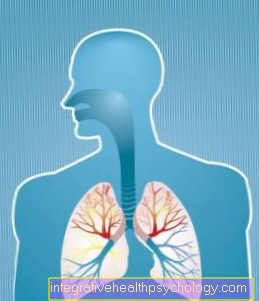
Like other organs, the eye is supplied with blood. To do this, an artery directs the oxygen-rich blood into the eye and a vein directs the oxygen-poor blood away again. In a stroke, the blood-draining vein becomes blocked. It is often closed by a thrombus. This is a collection of different components such as calcium and blood components. These thrombi can form on damaged arterial or venous walls in the blood system. They are the result of hardening of the arteries.
Patients with obesity, high blood pressure and cholesterol levels, and lack of exercise are among the high-risk patients who have more calcifications in the arteries. Even patients who have been working on one for a long time Diabetes mellitus can suffer from thrombi and injuries to the vessel walls due to increasing damage to the vessels.
There is a possibility that the thrombi will loosen and wash into other blood systems, including the eye. The stenosis (occlusion) can also have formed directly in the vein. Now the blood can no longer drain away and increasingly accumulates in the eye. The accumulated blood is ultimately also distributed to the individual layers of the eye. The retina is also affected. Finally, after a short time, the retina becomes very thick and bloody. Since the retina is important for vision, there is a risk that it will be injured by the increase in internal pressure in the eye caused by the massive congestion of blood. This can lead to a loss of vision, which can also become permanent (chronic) if prompt treatment is not taken.
Symptoms
The stroke in the eye often starts very suddenly and the patients usually do not notice the process at first. The vein is closed painlessly. Then suddenly various visual disturbances can occur after a stroke. The field of vision can be restricted, so that some areas are blurred or no longer visible. Typically, the visual field disorders are square in shape or half of the field of view is reduced. This also increases the risk of the patient falling. Impaired vision usually occurs when the vessel is only partially obstructed by a thrombus.
Read more about this at: Thrombosis in the eye such as These symptoms will help you identify a blood clot in your head
Furthermore, the patient can see blurred as if looking through a veil. These visual deficits are called scotomas in ophthalmology.
A different perception of the colors is also possible. The retina is restricted in its function by the accumulated blood. The thrombosis can also lead to a reduction in visual acuity, because the point of sharpest vision is the Macula, also swells.
However, if the vein is completely closed, the blood supply to the affected eye is interrupted. In this case, the patient is temporarily blind. This situation is very frightening for many people affected, because one is unsure where this symptom suddenly comes from and how to act. In this condition it is often difficult for the patient to read or recognize anything.
Many retinal vein occlusions occur at night, as the lying position increases the pressure in the veins and at the same time the arterial blood pressure is lower. The symptoms are often only noticed in the morning when the patient wakes up. In some cases, both eyes can be affected, but often only one side is affected. If visual disturbances occur and the cause is not yet known, the person affected should consult a doctor as soon as possible. Prompt treatment and exposure of the blood vessels is important, as consequential damage such as permanent visual impairment or, in the worst case, blindness can remain.
You can find more information about the signs of a general stroke on our website Signs of stroke or test for stroke symptoms - I can test this myself
Lightning bolts
The retina is very important for mapping what is seen. Without it, the information is not passed on to the visual center in the brain. Changes in the blood supply, such as is the case with a thrombosis and the subsequent stroke in the eye, are very sensitive and react immediately to them. The changed blood supply causes symptoms such as blurring of the objects, flashes of light and, finally, the complete vision can be lost and the patient only sees black in front of the eye.
In many diseases of the inner eye, flashes of light are the first warning signs of incipient damage to the retina (retina). For example, flashes of light occur not only with retinal vein occlusion, but also with diabetes as a result of vascular changes due to polyneuropathy and also with age-related vitreous changes.
In many cases, the retina becomes detached, which stimulates the sensory cells that are still in contact with the vitreous and thus generates the flashes of light. If flashes of light appear for the first time, the person affected should have their eyes checked by a specialist.
Typical signs of a stroke in the eye
Recognizing a stroke in the eye and acting in good time is not always very easy for the layperson. Especially since the symptoms can creep in and are often not noticed at first. The typical symptoms such as flashes of light, blurred or distorted field of view and partial visual field restrictions are typical signs of a stroke both in the eye and a stroke in a vessel of the brain.
Short-term blindness can also occur. The patient should be mindful of the first signs of change, which are also the first to occur. It is important not to let too much time pass before you see a doctor, as permanent damage can result. It is best if the person concerned see an ophthalmologist who can carry out an initial examination and thus also assess the situation. Another option is to drive to a hospital straight away.
Read more on the subject at: Symptoms of a stroke
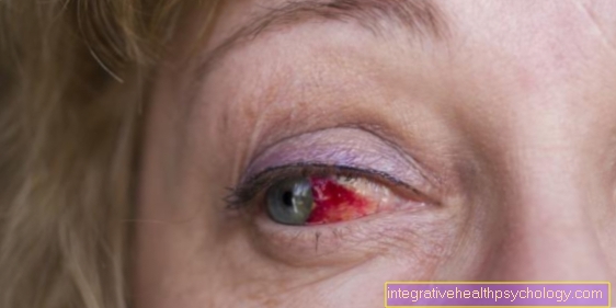
Broken vein in the eye - is that a stroke?
If you notice broken veins in your eye when you look in the mirror, this is initially no cause for concern. Numerous different causes can lead to this phenomenon. These include mechanical irritation from frequent rubbing or problems with contact lenses, but also dry eyes, foreign bodies or briefly increased pressure, such as when sneezing or coughing.
In almost all cases, broken veins in the eye are harmless and there is no need to think about the presence of a stroke. A specialist should only be consulted promptly if there are other serious symptoms, such as a visual impairment or the like.
Risk factors
There are several risk factors that can lead to stroke in the eye.
-
Artery occlusion:
-
Vascular inflammation, such as temporal arteritis
-
Atrial fibrillation
-
Atherosclerosis of the carotid artery (carotid stenosis)
-
-
Vein occlusion:
-
Diabetes mellitus
-
high blood pressure
-
arteriosclerosis
-
Green star (glaucoma)
-
Inflammation of the eye vessels (retinal vasculitis)
-
Diagnosis
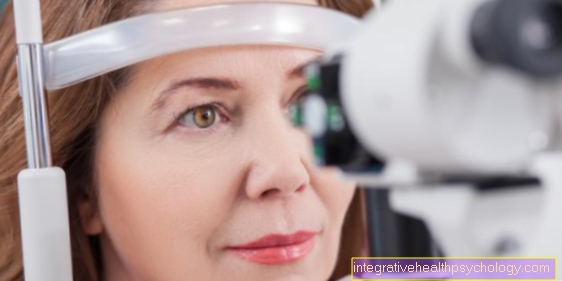
There are several tests that can be done to diagnose a stroke in the eye. The doctor can mirror the fundus. He looks at the retina with a special lamp. The first changes, such as swelling and blocked vessels, can be seen here. Since the retina can become detached or injured in the event of a stroke, it is important to determine the current condition of the retina. The subsequent treatment, which should also be carried out quickly, is geared towards this, so that no consequential damage such as loss of vision remains.
Using fluorescence angiography, the vessels of the retina can be better visualized and assessed. The intraocular pressure can also be measured. This examination is completely painless and provides information about the pressure inside the eye. By testing the visual acuity and the area of the field of vision, the doctor can also make initial assessments of the extent of the damage to the retina. The general functions of the eye are also checked. This includes mobility in all directions to check the optic nerve and also the pupil reflex. This is checked with a small lamp.
Also read: This is how a stroke is diagnosed
therapy
A early Treating a stroke is hugely important to Consequential damage like a permanent one Blindness of the affected eye prevent. The earlier the treatment, the better the chances. The focus is initially also on Preservation of eyesight. This is followed by the Fighting causes to minimize the risk of another stroke at the same time.
Different treatment approaches are carried out. It can blood-thinning drugs administered. This should allow the blood to flow better through the vessels of the retina. Since there is also a swelling in the eye, which hinders clear vision, kortisone-containing medicines administered. They are also injected directly into the eye under local anesthesia in the acute situation. They are supposed to cause the eye to swell better and the retina to suffer no damage.
Another very common remedy are VEGF inhibitors. This is a substance that is also injected directly into the eye. It prevents the growth of new vessels by inhibiting the growth factor VEGF. This prevents getting unwanted Vessels, the one Edema formation would also favor new education. At the same time, the inhibitors have a decongestant effect. In the long term, the cause, i.e. the poor quality of the vessels, should also be improved. So should a Treatment of high blood pressure and fat levels respectively.
Patient with a diabetes should be correctly adjusted and on a healthy nutrition respect, think highly of. Furthermore is also the Quit smoking and more physical activity conducive to the risk of a recurring stroke in the same or the other eye.
What to do?
Many people do not realize that a stroke can also occur in the eye. Since the symptoms can also occur gradually, the situation is initially not recognized until visual disturbances occur or even blindness in the affected eye. If the cause is not treated quickly, the retina can be severely damaged due to the accumulated blood. If the blood supply is interrupted for too long, it can be irreparably damaged. For the patient, this means that consequential damage such as partial visual impairment or complete loss of vision remains. Therefore, the person concerned should consult an ophthalmologist. For many, the complaints are unsettled and frightening. It is therefore helpful when the patient is not alone, especially since it would be too dangerous to drive a car in this situation.
consequences
The severity of the consequential damage caused by a stroke in the eye depends not only on the time it takes to initiate adequate therapy, but also on the vessel concerned. While occlusions of the lateral branch veins usually only lead to minor restrictions, the consequences of an occlusion of the central ocular vein can be considerable.
The most serious consequential damage is the blindness of the affected eye, which can be the case with delayed initiation of therapy.
Another early complication is the so-called macular edema. In this case, fluid escapes from the damaged vessels and collects in the macula, the place of sharpest vision. Most of the affected people then report a gray haze over the field of vision of the eye.
Long-term consequences of a vein occlusion in the eye are usually triggered by the formation of new vessels. The new vessels grow in uncontrollably and can thus cause damage. On the one hand, the rising tension can lead to the detachment of the retina, which can also be associated with considerable impairment of vision, but also to glaucoma.
The latter occurs when the newly formed vessels grow into the corner of the chamber and the eye fluid can no longer drain away. The result is increased intraocular pressure which, if it persists for a long time, can damage the optic nerve.
The stroke can lead to further important consequential damage. Find out more at: These are the consequences of a stroke!
blindness
Occlusions in the arterial and venous supply to the retina are among the most common causes of blindness. While arterial occlusions in particular lead to a loss of vision in the acute phase, the long-term consequences of both forms, such as the formation of new vessels and detachment of the retina, are the most common causes of blindness due to a stroke in the eye.
Therefore, in addition to acute therapy, the eye must also be closely checked by a specialist during the course of the treatment so that these complications can be recognized as early as possible and thus treated.
Eye flicker
The eye flicker mostly affects the outer field of vision and is described as the moving back and forth of small bright points. This can lead to impaired eyesight. The causes of eye flicker can be diverse and range from migraines to circulatory disorders.
However, suspicion of a stroke in the eye should be clarified, particularly if this symptom persists. After therapy of an underlying circulatory disorder, the eye flicker disappears again.
Read more: Eye flicker
Double vision
Double vision can be caused by a number of different causes. What these causes have in common is that they cause restricted and asynchronous movement of the eye muscles.
In addition to paralysis of the eye muscles or inflammation of the brain, strokes are also often the trigger for this symptom. In most cases, however, double vision suggest a circulatory disorder in the brain stem. The supplying vessels of the eye muscles are rarely affected.