SLAP lesion
introduction

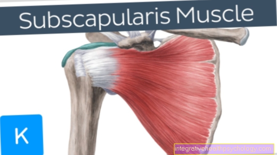
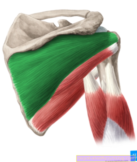
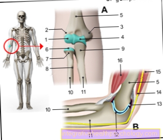
The Shoulder joint consists of the Swivel head, which is part of the humerus head and which Socketthat are between shoulder blade and Collarbone is located.
The joint socket is smaller than the joint head and for this reason does not offer the necessary stability to support the upper arm to keep safely in the pan.
For this reason, the joint is stabilized on the one hand by muscles that stretch around the upper arm and shoulder joint, and on the other hand by the so-called labrum.
The labrum is a joint lip that wraps around the socket, giving the socket the necessary enlargement.
The advantage is that the joint lip does not have a solid structure such as e.g. has a bone and so the upper arm has sufficient freedom of movement in the pan.
A muscle tendon is attached to the upper part of the joint lip and Biceps muscle is expected. Both structures are anatomically also called Labrum-biceps complex designated.
Injuries and damage to this complex are considered Slap lesion designated.
Appointment with a shoulder specialist

I would be happy to advise you!
Who am I?
My name is Carmen Heinz. I am a specialist in orthopedics and trauma surgery in the specialist team of .
The shoulder joint is one of the most complicated joints in the human body.
The treatment of the shoulder (rotator cuff, impingement syndrome, calcified shoulder (tendinosis calcarea, biceps tendon, etc.) therefore requires a lot of experience.
I treat a wide variety of shoulder diseases in a conservative way.
The aim of any therapy is treatment with full recovery without surgery.
Which therapy achieves the best results in the long term can only be determined after looking at all of the information (Examination, X-ray, ultrasound, MRI, etc.) be assessed.
You can find me in:
- - your orthopedic surgeon
14
Directly to the online appointment arrangement
Unfortunately, it is currently only possible to make an appointment with private health insurers. I hope for your understanding!
You can find more information about myself at Carmen Heinz.
Cause of the SLAP lesion
The reason why a SLAP lesion triggered, can be acute or chronic in nature.
One of the chronic causes is overload in the area of the Shoulder joint.
If excessively large loads are carried or balanced or lifted over a long period of time, the entire Shoulder joint including the labrumbiceps Complex things are under so much stress that at some point a crack or tear will result.
In addition to chronic overstrains, chronic inappropriate strains can also lead to parts of the labrumbiceps Complex are more heavily loaded than other parts. This can also lead to cracks or tears.
Some sports are also always popular as risk factors for slap lesions. As a rule, these are racket-swinging sports such as baseball, tennis or table tennis, which, due to the repetitive arm movement, place particularly high stress on the area shoulder to lead.
Are very heavy loads carried acutely (E.g. lifting weights for untrained people), there may also be immediate cracks or tears.
The same thing can happen in an accident. So-called high-speed trauma, such as those that occur in car accidents or in sports accidents, can trigger an acute slap lesion. If the shoulder is squeezed or twisted unchecked, these acute slap lesions can occur.
Symptoms
Is it a chronic one? Slap lesion, the patient may not notice anything at first.
With advancing and untreated lesion, the patient is mostly exposed to heavy loads Pain indicate, while acute slap lesions or advanced lesions indicate immediate pain.
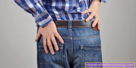
The pain character is called biting or burning described, it is localized in the shoulder area and can also extend over the entire shoulder to the upper back.
The pain means that the patient often goes into relieving postures in order to reduce the severity of the pain. These postures usually have the consequence that the Shoulder joint Then the stress is often incorrect, which in turn can lead to muscle hardening, bone wear and tear and further pain.
In addition to the pain that occurs even at rest when the slap lesion has progressed, movement impairments can also occur, particularly in the case of severe courses. On the one hand, these come about because the patient no longer fully performs the movements due to the pain.
Another and perhaps more important reason in this context is a resulting instability in the Shoulder jointwhich is due to the fact that the shoulder joint stabilizing effect due to the tear or tear of the labrum-biceps complex is no longer or only insufficient.
Sometimes this instability can be so severe that the patient can only raise the arm up to an angle of 90 degrees and then stop moving.
The fear of one Dislocation of the joint also means that the patient is very careful in arm movements and is accordingly restricted in his everyday life.
Diagnosis of a SLAP lesion
Diagnosing a Slap lesion it's not always easy. In any case, it is important to conduct a detailed patient survey, which should provide information on whether the patient is one of the risk patients for a slap lesion (Incorrect loads, overloads, athletes of racket or ball sports) heard.
Then you should ask exactly which symptoms occur and, above all, during which movement.
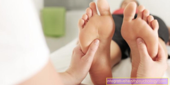
After the interview, a detailed physical examination should be carried out, which initially involves active movement in the Shoulder joint should explore (what can the patient do himself?), then a passive movement by the examiner (the patient lets his arm hang down, the movements are carried out by the examiner).
If this hardens the suspicion of a slap lesion, e.g. If instability is detected, it should be considered which imaging method should be used.
Either Ultrasonic as well as the classic roentgen can represent the shoulder joint but are very limited in terms of the representation of the soft tissues in the joint. Both examination methods cannot or hardly show a slap lesion.
Often times, the SLAP lesion can only MRI of the shoulder from (magnetic resonance imaging / shoulder), which can give a clearer, if not one hundred percent insight, be made visible.
But even in the MRI of the shoulder, this often cannot be detected without contrast agent that has to be injected directly into the joint. Since the injured biceps tendon anchor is very small, it is often not possible to reliably classify the SLAP lesion in an MRI of the shoulder, even with contrast media.
The safest way to prove a slap lesion today is this Jointoscopy. It is usually only carried out if previous examinations have provided no evidence of a slap lesion, but the symptoms of a slap lesion are typical.
With the also as Arthroscopy For the designated examination, two small skin incisions are made on the disinfected shoulder joint and the camera and examination equipment are inserted into the joint.
The camera delivers live images and sends them to a monitor. With the help of the second instrument, straightening devices, scissors and pliers can be inserted into the joint.
treatment
With a manifest Slap lesion The surgical treatment method is often the only therapeutically sensible approach.
Sometimes it is that diagnostic mentioned above Arthroscopy is already being used for therapeutic treatment. Torn parts that are seen during the examination are tied back together with sutures.
Free tissue that is created in the case of tears and is located in the joint space and also potentially interferes with the movement in the joint is usually grasped with the help of small forceps and removed from the joint.
Depending on the severity of the slap lesion, the procedure takes a few minutes up to 1-2 hours.
Today, arthroscopic treatment is rarely started and open shoulder surgery is continued. This can happen if the examiner has anatomical features of the Shoulder joint cannot get a proper insight or cannot treat the corresponding lesion due to lack of space.
After therapeutic treatment of a slap lesion, the arm can usually be fully loaded immediately. However, it must be ensured that movements that have led to this injury should initially not be carried out. Physiotherapy treatment can also be useful. Treatment of pain is usually no longer necessary after the procedure. If so, cooling measures or anti-inflammatory pain relievers should be given.