Stages of osteochondrosis dissecans
introduction
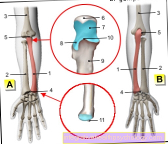
In the Osteochondrosis dissecans part of the bone dies beneath the articular cartilage so that it can peel off and is loosely present in the joint cavity (Dissectate). The exact causesthat lead to this disease are still not fully understood, however, repetitive trauma or overuse as well as circulatory disorders in the bones seem to play a role. Infections of the bone, however, play no role in the development of the Osteochondrosis dissecans. The disease is thus an aseptic bone necrosis.

Osteochondrosis can occur on joints all over the body or, in children, on growth plates.
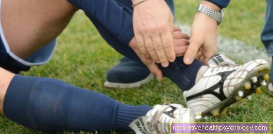
- Osteochondrosis dissecans on the ankle
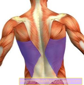
- Osteochondrosis dissecans on the knee
If osteochondrosis dissecans is diagnosed on x-rays or an MRI, it is important to determine what stage it is at, as treatment for osteochondrosis dissecans varies depending on the stage.
Stage 1
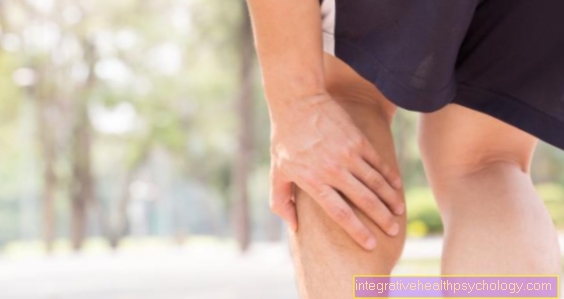
In the first stage, the bone begins due to unknown processes to become lytic, that is, it begins to dissolve. in the X-ray image becomes this stage mostly not visiblebecause the bone is not interrupted at any point. If a MRI of the knee joint is made, you can in this one small osteolyses as foci that distinguish themselves from normal bone density. There is also a Bone marrow edema, a small accumulation of fluid in the bone tissue that appears as a lightening in a water-weighted sequence of the MRI. Of the Cartilage is still intact and fulfills its mechanical function in the joint, hence are Symptoms rather weak. You may experience slight pain under exertion.
At this stage the Therapy of choice is conservative to choose. The further course of the disease is observed; there may be phases in which the affected joint should be spared from heavy stress.
Appointment with a knee specialist?
I would be happy to advise you!
Who am I?
My name is I am a specialist in orthopedics and the founder of .
Various television programs and print media report regularly about my work. On HR television you can see me every 6 weeks live on "Hallo Hessen".
But now enough is indicated ;-)
The knee joint is one of the joints with the greatest stress.
Therefore, the treatment of the knee joint (e.g. meniscus tear, cartilage damage, cruciate ligament damage, runner's knee, etc.) requires a lot of experience.
I treat a wide variety of knee diseases in a conservative way.
The aim of any treatment is treatment without surgery.
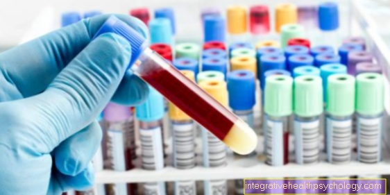
Which therapy achieves the best results in the long term can only be determined after looking at all of the information (Examination, X-ray, ultrasound, MRI, etc.) be assessed.
You can find me in:
- - your orthopedic surgeon
14
Directly to the online appointment arrangement
Unfortunately, it is currently only possible to make an appointment with private health insurers. I hope for your understanding!
Further information about myself can be found at
Stage 2
When the disease continues to progress, the bone is increasingly broken up into several fragments. The necrosis of the bone becomes larger as it is numerous Trabeculae broken down become or collapse. In this case you can go from Microfractures speak.
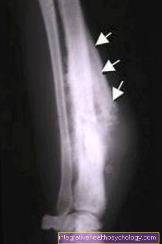
At the same time, the body reacts to the breakdown by Initiates processes for stabilization. The damaged areas below the cartilage are covered by a Sclerotherapy reinforced. This means that more bone tissue is stored in this area. in the X-ray image one sees this sclerosis as lighter line below the articular cartilage.
At this point the risk of further damage is greater than in stage 1. To prevent damage to the cartilage, a Sports ban imposed to give the body time to regenerate the defects. Especially with young people Until puberty, the growth plates of which are still open, there is a good one Self-healing tendency.
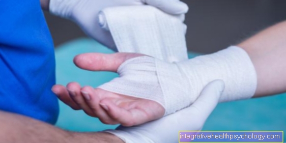
Forearm crutches can be prescribed for support. if the Pain symptoms gets worse despite a break in sport, the indication for Jointoscopy posed. The Cartilage assessed become. In stage 2 it is still intact. The doctor can then drill into the bone through the cartilage and stimulate the tissue to develop further.
Stage 3
The next step is to Fragmentation. This is traditionally referred to when ball joints such as the femoral head are affected, as this ball disintegrates in the X-ray image and small fragments remain. It also occurs in other affected joints first detachment of the cartilage-bone structure. First of all, the detached fragment remains (Dissectate) but in connection with its document. As part of a Jointoscopy the examiner can reach under the piece of cartilage with a small hook and lift it, but it is not yet detached.
Both in the X-ray image, but especially in the MRI one sees one Interruption of the normally smooth joint space. In the MRI it becomes clear that the interruption took place in the area of the cartilage. Sclerosis and edema (fluid margin) are still visible. The Pain symptoms increase significantly. In some cases, acute pain when moving can occur when the damaged cartilage becomes trapped.
As part of the Knee reflection can the Piece of cartilage fixed again become. Especially in young patients and with more superficial defects, there is a good chance that the cartilage will grow solid again.
Stage 4
The Dissectate can peel off and then lies as a free body in the joint cavity. On the side from which the dissectate has detached, you can see an empty "dissectate bed". The free dissectate can move when moving and so substantial Pain caused by an entrapment.
In this case too, a Jointoscopy carried out. If the cartilage still appears vital despite detachment, it can be fixed again with small pins, as in stage 3. If the defect is very deep or the piece of cartilage is obviously dead, the joint mouse is removed and the dissection bed filled.
Either a Cartilaginous cylinders taken from a less stressed area of the joint and transplanted into the defect zone or one Cartilage cell transplant carried out. Either new cartilage is grown from the body's own cells, which takes a long time, or, alternatively, a prepared collagen fleece is placed in the defect. However, in contrast to the cartilage-bone cylinder, these procedures only produce good results in the case of superficial defects.
forecast
Overall can Healing is achieved without restriction at any stage become. It is important to note, however, that cartilage is a slowly regenerating tissue. It follows that it Last months to years can until a completely unrestricted use of the joint is possible.