Venules
introduction
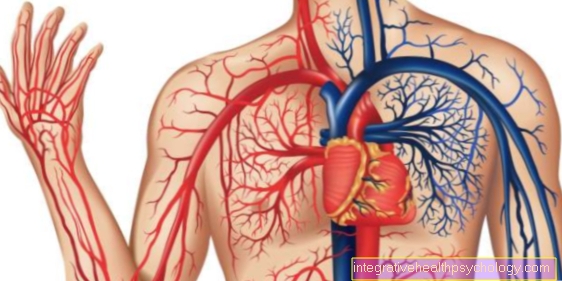
The term venole describes a vascular section in the body's blood vessel system which, together with the arterioles and capillaries, forms the end flow path of the vascular system.
The role of the venules includes the exchange between blood and tissue and the transport of the blood as part of the vascular system. It collects and directs the blood from the venous part of the capillary bed and flows together with other venules, which eventually form a vein.
The venole differs from the veins both in its wall structure and in its function.
There is a special form of venules in the lymphoid tissue. These enable the lymphoid cellsthat are temporarily in the blood migrate back into the lymphatic tissue. The venules have a particularly permeable wall and thus ensure the exchange of tissue and blood.

anatomy
The large transport vessels of the body are basically made of three wall layers, the Tunica intima, the Tunica media and the Tunica adventitia. These, in turn, contain some sub-layers that vary in strength depending on the location and function of the vessel.
The Tunica intima consists mainly of so-called Endotheliumwhich for the Mass transfer responsible for. It also contains connective tissue.
In contrast, the Tunica media from smooth annular muscles and elastic fibers, which serve as muscle pumps for the blood vessel and are essential for blood transport.
The Tunica adventitia is the outer layer of the vessel and is exposed loose connective tissue together. This layer stabilizes the vessel in the surrounding tissue and can also contain blood or lymph vessels and nerve tracts.
In contrast to the large vessels, the small venules no or only a very thin tunica media. This layer gives the vessel wall stability. Since the main function of the venules in the Exchange of nutrients with the surrounding tissue, this wall layer is not required.The part of the venole facing the capillary bed therefore does not contain a tunica media. In the course of the venules, a thin layer of smooth muscle is created.
The blood pressure in the venules is only very low, so that the wall layer of tunica intima and adventita is sufficient. The tunica media would only represent a barrier to the exchange of substances. In addition, the venules possess no venous valves compared to the veins. Venous valves have a valve function in the large body veins and facilitate the return of blood to the heart by preventing the blood from flowing back.
Difference Between a Venole and an Arteriole
A Arteriole is also a component of the terminal vascular system and resembles an artery in its wall structure.
The arteries generally have a larger and more compact muscle layer than the veins. The arterioles form the resistance vessels in the body's circulation and therefore have a significantly thicker muscle layer than the venules. The wall layer made of smooth muscle cells is used to Regulation of blood pressure in the subsequent capillaries. The upstream arteries conduct the blood with high blood pressure. This is strongly throttled in the area of the arterioles and for each subsequent one Organ individually adapted.
If there is a great deal of blood loss, for example, they can reduce the blood flow to the end current path so that the remaining blood is centralized. This function is essential for maintaining cardiovascular stabilization.
What is a shunt?
At a Shunt it is an arteriovenous anastomosis, i.e. one direct transition from an arteriole to a venole, without an intermediate capillary bed. The blood flow to the neighboring capillary beds can be regulated through these shunt connections. So can some Capillary areas completely switched off if necessary become. The blood flow is also regulated by the wall muscles of the arterioles.
A shock can lead to a loss of regulation of the capillary blood flow. As a result, there is more blood in the capillaries and the central vessels and the heart lacks this, so that circulatory failure can occur.


.jpg)


