Baker's cyst
Synonyms
- Popiteal cyst
- Synovial cyst
- Bulging of the joint capsule
- Popliteal cyst
Definition of Baker's cyst
A Baker's cyst is caused by a Knee joint disease with chronic knee joint effusion. This creates a bulging (protuberance) of the posterior joint capsule, comparable to an overflow valve.
Alternatively, mechanical irritation of the muscles in the hollow of the knee can result in ganglia (cavities filled with gelatin) that are deposited in the hollow of the knee.
A Baker's cyst is particularly common in older people due to wear and tear of the knee joint and in children (usually without a clear cause).

General
The so-called Baker's cyst is a sac-shaped, fluid-filled sac in the central (medial) hollow of the knee. Its name comes from its first describer W. M. Baker, an English surgeon from London in the 19th century.
The Baker cyst always starts from the knee joint capsule. From the joint capsule it is connected to the main chamber via a narrow web or passage (stem-like connection). In the case of a Baker's cyst, which is located in a typical position, the connecting duct pushes through the muscular structures of the gastrocnemius muscle (head mediale) and the semimembranosus muscle (flexor muscles of the thigh). If a Baker cyst persists for a long time, several cyst chambers can form, which makes puncturing the cyst particularly difficult.
Read more on the topic: Baker's cyst in the knee
Formation of a Baker's cyst
A Baker cyst is often the result of an internal knee disease. In the context of rheumatoid arthritis (rheumatism) or chronic meniscus damage, permanent joint effusion occurs (water in the knee joint). The associated chronic increase in internal joint pressure ensures increasing fatigue and slackening of the joint capsule of the knee joint and can lead to permanent bulging of the capsule and thus to the formation of a poplietal cyst = Baker's cyst.
Because of its swelling nature, a Baker cyst can resemble a tumor of the back of the knee, so that a malignant disease must always be excluded. However, this can easily be achieved with a sonographic examination of the hollow of the knee. If the cyst ruptures / ruptures as a result of increasing the chamber pressure, i.e. a rupture with leakage of fluid into the tissue, swelling in the affected area and pain that increases with pressure can be found. This condition can easily be mistaken for deep vein thrombosis. However, if the cause is not eliminated, the Baker cyst will reappear, with more ventricular formation.
Appointment with a knee specialist?
I would be happy to advise you!
Who am I?
My name is I am a specialist in orthopedics and the founder of .
Various television programs and print media report regularly about my work. On HR television you can see me every 6 weeks live on "Hallo Hessen".
But now enough is indicated ;-)
The knee joint is one of the joints with the greatest stress.
Therefore, the treatment of the knee joint (e.g. meniscus tear, cartilage damage, cruciate ligament damage, runner's knee, etc.) requires a lot of experience.
I treat a wide variety of knee diseases in a conservative way.
The aim of any treatment is treatment without surgery.
Which therapy achieves the best results in the long term can only be determined after looking at all of the information (Examination, X-ray, ultrasound, MRI, etc.) be assessed.
You can find me in:
- - your orthopedic surgeon
14
Directly to the online appointment arrangement
Unfortunately, it is currently only possible to make an appointment with private health insurers. I hope for your understanding!
Further information about myself can be found at
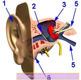
Illustration of a Baker's cyst

- Baker's cyst
(Popliteal cyst) - Femur -
Femur - Semi-membranous muscle -
Semimembranosus muscle - Joint capsule, fiber layer
(yellow) -
Capsula articularis,
Membrana fibrosa - Joint capsule, soft layer
(orange) -
Capsula articularis,
Synovial membrane - Joint cavity
(filled with synovial fluid) -
Articular cavity, synovia - Shin - Tibia
- Internal calf muscle -
M. gastrocnemius, caput mediale - Fibula - Fibula
- Articular cartilage (dark blue) -
Cartilago articularis - Kneecap - patella
- Inner meniscus -
Meniscus medialis
A - Knee joint effusion with Baker's cyst
B - Healthy knee joint
a - swelling in the hollow of the knee
b - swelling in the calf muscles
You can find an overview of all Dr-Gumpert images at: medical illustrations
MRI knee joint with Baker's cyst

(left kneecap, right hollow of the knee)
- Thigh (femur)
- Shinbone (tibia)
- Baker's cyst (Politeal cyst)
- meniscus
root cause
The root cause for training a Baker's cyst in the Hollow of the knee is the increased production of Synovial fluid in the knee joint. The cause of this, in turn, is usually damage to the knee joint, such as a arthrosis, a longer one Meniscus damage or an underlying inflammatory disease such as a Rheumatoid arthritis. Responsible for training a Baker's cyst is in the majority of cases a wear-related cause, i.e. osteoarthritis or a meniscus tear. The knee joint tries to improve the function of the knee joint by increasing the production of “synovial fluid”. This leads to a permanent increase in internal joint pressure. The weakest point of the knee joint capsule gradually gives way and forms the Baker's cyst. This forms an “overflow bag”, which is connected to the knee joint via a stick-shaped connection. This stalk almost always runs between the central (medial) head of the Gastrocnemius muscle (Calf muscle) and the insertion tendon plate of the flexor muscles (Semimembranosus = muscle Thigh muscle). On palpation, a closed connective tissue capsule filled with fluid can be felt in the back of the hollow of the knee, which is particularly difficult with small cysts. For such cases is the Ultrasound examination (Sonography) groundbreaking.
frequency
A Baker's cyst can in principle occur at any age. However, children are less affected by the development of cystic changes in the hollow of the knee. If a Baker cyst does develop in childhood, boys are affected twice as often as girls. (see also: Childhood Baker's cyst)
The disease can predominantly be observed in middle and advanced age (typically 60 years and older).
Symptoms
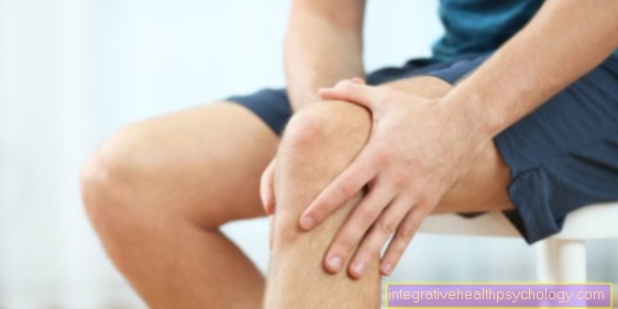
Patients with Baker's cyst report recurrent knee and upper calf pain that can be located on the back of the leg. In some cases, only an uncharacteristic feeling of tension in the hollow of the knee is reported. The extent of the discomfort, however, depends on the level of activity of the fluid formation. After exercise, a Baker's cyst typically swells a lot and can no longer be detectable after a few days with care. Accordingly, the symptoms usually fluctuate with the degree of stress, corresponding to the tension of the fluid-filled cyst.
Once the Baker cyst has reached a certain size, the pain can also occur completely independent of the load.
More on this topic can be found here:
- Baker's cyst symptoms
- Acute knee pain - that may be behind it
Symptoms of a burst Baker's cyst

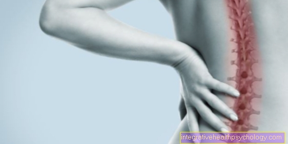
In most cases, affected people feel a feeling of pressure in the hollow of their knees. Depending on the size of the Baker cyst, from approx. 2 cm, it can also be easily felt in the hollow of the knee. The symptoms also depend on physical activity. The more active the person is, the clearer and stronger the symptoms are. Pain can occur especially when bending the knee. In addition, strong pain can be provoked by applying pressure to the hollow of the knee. Depending on the size and displacement of the surrounding tissue, Baker's cysts can simulate lower leg thrombosis by compressing blood vessels. If the cyst compresses nerves in the hollow of the knee, sensory disturbances such as numbness or paralysis can occur in the lower leg or foot. In addition to the tactile findings, ultrasound or magnetic resonance imaging can confirm the diagnosis.
If the Baker's cyst ruptures as the symptoms progress, the fluid from the cyst can spread into the surrounding tissue and the pain worsens. In addition, the fluid from the cyst can spread into the lower leg muscles. This causes a strong inflammatory response, which is accompanied by severe pain and swelling. These are symptoms that can be similar to deep vein thrombosis. That is why it is important to rule out differential diagnoses such as thrombosis during a closer examination.
diagnosis
The diagnosis of a Baker's cyst can generally be made relatively easily from the combination of medical history, complaints, clinical and diagnostic examination, provided the doctor thinks of this possibility of diagnosis. In the case of pronounced forms, the Baker's cyst bulges out in the hollow of the knee, medium-sized sizes can usually be felt in a classic location, small Baker's cysts can usually only be visualized with diagnostic methods.
The following diagnostic methods can help to confirm the diagnosis:
- An ultrasound examination (sonography) of the hollow of the knee can detect a Baker cyst and show its location and dimensions.
- Conventional X-rays can show arthritic changes (wear and tear as the cause of the cyst) in the knee joint.
- A magnetic resonance tomography (MRT) of the knee can also show the exact anatomical location and the connection to the joint capsule. Occasionally Baker's cysts cause diagnostic problems due to their unusually bizarre, also very long tubular configurations in the case of bleeding with and without ruptures. An MRI is certainly not necessary for the detection of a classic Baker cyst. However, since the cause of a Bakerzste must always be treated, the MRI provides helpful additional information about accompanying injuries, such as meniscus tears or the degree of osteoarthritis.
- Thigh (femur)
- Kneecap (patella)
- Knee joint (articulatio genu)
- Shinbone (tibia)
- Baker's cyst (Politeal cyst)
therapy

Physiotherapy is a common form of therapy for Baker's cysts. If the Baker's cyst does not cause any symptoms, then it does not necessarily have to be treatment respectively. If the mobility of the knee is restricted by the cyst or if pain occurs, there is the possibility of one conservative therapy and on the other hand one surgical intervention to undergo. It depends on the extent of the swelling and the symptoms it causes.
If symptoms occur, anti-inflammatory drugs such as Diclofenac or Ibuprofen are given. In very acute, painful cases, a Cortisone syringe be helpful. The method with cortisone is not the first choice because of the side effects.
Furthermore, additional measures such as physical therapy or Physical therapies help improve symptoms. It is important, however, that the cause of the Baker's cyst is often an underlying disease of the knee joint that is chronic Joint effusions goes along like for example Cartilage- or Meniscus damage. For this reason, it is essential to repair the causal damage. If this underlying disease is successfully treated, a Baker's cyst can regress on its own in the course of the therapy.
More on this topic can be found here: Baker's cyst treatment
OP of a Baker's cyst
If the Baker's cyst increases in size and functional impairment due to disruption of surrounding structures such as Blood vessels and annoy surgery should be considered. The Baker's cyst is surgically removed. Firstly, the cyst can be surgically removed by making an incision in the Hollow of the knee sets, the cyst is freed from surrounding structures and completely removed after constriction at the stem of the cyst. Then the Joint capsule locked. This is the way to try Recurrences to avoid.
There is also the option of having the cyst punctured and sucking off the fluid. With this approach, however, it is likely that the cyst will come back, i.e. a relapse will occur.
In addition, the underlying disease should be corrected before the cyst is removed, because otherwise there is also the possibility that the Baker cyst will recur.
More on this topic can be found here: Baker's cyst surgery
Puncture of a Baker's cyst
The Puncture represents one important part of the therapy options a Baker's cyst. The attending physician performs a Insert a needle into the cyst and away with this one in the cyst contained liquid. In addition to a purely conservative or surgical therapy, the puncture represents a kind of compromise between these strategies. However, it should be noted that a Puncture only treat symptoms represents a Baker's cyst and the inflammation, and thus the cause of the formation of the cyst, can not handle.
Because of this, be often other therapies with the puncture of a Baker's cyst combined to achieve the best possible treatment success. target of these therapies is to one To prevent refilling of the cyst. For example anti-inflammatory drugs be taken, or the cortisone washes around the empty cyst become. A bandage which is wrapped around the knee joint can also contribute to the success of the treatment. The Puncture is a Baker's cyst not without complications and should be because of this only after an intensive medical consultation be performed. If the puncture is unsuccessful, surgical removal of the cyst can be considered.
More on this topic can be found here: Baker's cyst puncture
Homeopathy for a Baker's cyst
In addition to a large number of available therapy options to treat a Baker's cyst is the Taking homeopathic remedies a popular attempt among those affected to treat the cyst as a self-treatment. It should be noted that the therapy of Baker's cyst with homeopathic remedies Not recommended from a medical point of view can be as it no hints insists that the homeopathic remedies available are a healing effect have on the cyst. So should always be a Advice from the attending physician which can explain the individual therapy options and confirm the safe use of homeopathic remedies, possibly with simultaneous conservative or surgical therapy.
When taking homeopathic remedies, complications are usually not to be expected. It should be noted, however, that if therapy is not received, disease progression and the inflammation associated with Baker's cyst are possible and subsequent treatment may be associated with complications.
Taping a Baker's cyst
The Applying a tape bandage is very popular for complaints of the knee joint. The elastic tape can help to increase the stability of the knee joint and to reduce pain during exercise, especially when it comes to complaints of the muscles or ligaments. The Baker's cyst is usually based on an inflammatory process of the knee joint and can be Do not treat successfully with the application of an elastic tape dressing alone. Nevertheless, tape bandages can be one meaningful addition to the therapy options a Baker's cyst. Whether the use of a kinesio tape bandage makes sense in individual cases can be discussed with the treating doctor or physiotherapist.
Especially if the Baker's cyst has been removed with a puncture or surgery, a tape dressing can prevent a Baker's cyst from recurring. In addition to taking anti-inflammatory drugs and classic physiotherapy with lymphatic drainage, the bandage can be a useful measure of aftercare.
forecast
Conservative measures usually only lead to an improvement in the symptoms caused by the Baker's cyst. A disappearance or "drying up" of the Baker cyst is not to be expected when purely conservative measures are used. Only surgical therapy of the cause of the excessive water formation in the knee joint (e.g. a Meniscus damage) leads to the disappearance of the Baker cyst without operating on the Baker cyst itself. A direct Baker's cyst operation leads to the complete removal of the cyst, however, a high recurrence rate must be expected after an operation as long as the cause in the knee joint has not been eliminated.
Exercise for a Baker's cyst
The formation of a Baker's cyst is a relatively widespread disease in the population.After diagnosing a cyst, many affected people wonder whether something speaks against exercise while they have a Baker's cyst. A general recommendation is difficult to make become. A Baker's cyst usually develops due to an inflammatory process in the knee joint. A increased stress on the joint can this stimulate inflammatory processes and therefore can not recommended. This is especially true for large cysts and then if Pain occur. In particular from the Performing certain sports, which with a high stress on the knee must therefore go hand in hand (for example jogging or certain ball sports) with an acutely occurring Baker's cyst advised against become.
Sports which only one low load of the knee joint are, however in certain circumstances easily feasible. Depending on the type of movement, physical activity can even have a positive effect on the success of a Baker's cyst. Exerciseswhich by a treating Physiotherapist recommended can be used for conservative treatment of Baker's cyst and surgical follow-up care promote rapid healing of the cyst.
After operational care the cyst should no heavy load for some time be exercised on the affected knee joint. The attending surgeon can best provide information about the length of the sports break.
Baker's cyst in a child
The Baker's cyst can occur at any age, so a Baker's cyst can also develop spontaneously in the knee joint of a child.
The cause of one Baker's cyst in a child is still unexplained, but one assumes a congenital weakness of the joint capsule, which is why it can protrude towards the hollow of the knee if the pressure in the knee joint is too high.The children notice a feeling of tension in the joint more than a direct one Pain in the knee. A is also recommended for children Ultrasonic or one MRITo perform an examination of the knee joint. Differential diagnoses such as Hematomas or even Tumors of the bones or soft tissues are excluded.
There is often one in childhood spontaneous reduction in cyst size up to the complete disappearance of the cyst. For this reason, it is recommended to wait and see whether the cyst disappears on its own. In the case of extreme swelling with restrictions in the knee joint, a puncture of the cyst can be considered. Surgical removal, on the other hand, is rarely induced.
It is important for all parents: clarify the cause -> if no cause of the Baker's cyst can be found in the child -> keep calm!Read more detailed information on the topic at: Baker's cyst in a child
Summary
The Baker's cyst (Poplietal cyst) in the hollow of the knee is a fluid-filled protrusion of the hollow of the knee. It is an indirect sign of damage to the knee joint.
Due to the damage in the knee joint (causes for this can be wear and tear, ie arthrosis, Meniscus damage or an underlying inflammatory disease such as rheumatism or Rheumatoid arthritis his) there is an increased Water formation in the knee. The knee joint tries to “lubricate” the knee better, but produces too much “synovial fluid” overall. Due to the permanently increased internal joint pressure due to increased synovial fluid, the surrounding connective tissue slackens and a fluid-filled cyst forms. The weakest point of the joint capsule gives way, typically the inner hollow of the knee, and forms an "overflow sac" between two muscles.
A Baker's cyst is a disease of middle to old age (due to increasing wear and tear), although children can also be affected occasionally.
Pain as well as a movement-dependent often recurring Swelling in the back of the knee and the upper calf muscles are the main features. If the cyst breaks (ruptures), patients report severe sudden onset Pain in the back of the knee. If the cyst has a large volume, swelling in the area of the ankle can briefly occur. In most cases, if the Baker cyst ruptures, it can be detected again. - Thigh (femur)


.jpg)