Tooth pulp (pulp)
introduction
The anatomy of the tooth consists essentially of three layers. In the crown area, the outermost layer is the tooth enamel, the hardest substance in the body. This is followed by the dentin or dentin and inside is the pulp (pulp). The tooth root surrounds a third hard tooth substance, called cement, as the outermost layer, which serves to anchor the tooth and is therefore counted as part of the tooth holding apparatus. Then follows the dentin and inside the root canal with the root pulp.
Structure of the pulp
.jpg)
The pulp fills the inner cavities of the tooth. It adapts to the shape of the dentin (Dentine) roughly on. A distinction is made between the crown pulp and the root pulp. The pulp is well protected by the dentin and the enamel. In young people, the pulp cavity and root canals are initially very spacious. With age, both become more and more due to sustained dentine production (Secondary dentist) narrowed.
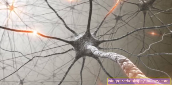
The internal structure of the tooth pulp consists of connective tissue, blood vessels and nerve fibers. At the edge of the pulp there is a layer of odontoblasts, cells that form new dentin and thus narrow the cavity. The pulp is supplied with blood by supplying and draining blood vessels through the opening at the tip of the root. This opening in the apex of the root also supplies nerve cells from a cranial nerve known as the trigeminal nerve. The pulp is connected to the entire organism through the opening at the tip of the root.
You might also be interested in: Pulp necrosis
Pulp diseases
_2.jpg)
The pulp can become ill through various influences. An inflammatory reaction of the pulp occurs predominantly as a result of progressive caries. But thermal stimuli, such as heating from grinding the tooth or chemical and toxic stimuli from tooth filling, can lead to a reaction in the pulp. Even through the opening at the tip of the root, the pulp can become inflamed in very deep periodontal processes.
The inflammatory reactions of the pulp can take place in different stages. Initially, only the crown pulp can be affected and then spread to the entire pulp. In the further course the pulp can either necrotici.e. may be dead or turn into a purulent disintegration of the pulp tissue, which is called gangrene. Since an inflammation is always accompanied by edema, there is great pain, as the inflammatory tissue in the pulp cavity cannot expand and therefore presses on the nerve fibers. The pain is therefore the main symptom of inflammation of the pulp.
Occasionally, a so-called denticle can cause pain. This is a hard structure with a dentin-like structure that is free or attached to the pulp wall within the pulp cavity. Dental diagnosis can mainly be made by x-rays.
Tooth pulp inflammation
A Pulpitis (Tooth pulp inflammation) is a disease that is characterized by the occurrence of inflammatory processes within the dental pulp.
The main reasons for the development of tooth pulp inflammation are mechanical, thermal and chemical irritation.
Metabolic products of bacteria, depth carious defects and / or cracks in the tooth structure can lead to a Pulpitis to lead.
Most of those affected complain of strong, stabbing symptoms in the course of this disease Toothachethat occur mainly when eating and drinking.
Short-term pulpitis that has a chance to heal is shown as a brief pain that is confined to one tooth.
The chronic one Pulpitis however, it becomes noticeable through permanent toothache and urgently needs to be treated by a dentist. Such a dental disease usually proceeds according to the same pattern, it begins with an inflammation in a limited area of the dental pulp (partial pulpitis).
When not treating a Tooth pulp inflammation the inflammatory processes continue into the pulp in the area of the crown (Pulp cavum) and then penetrates into the root canal. In the course of the inflammatory processes, so-called Endotoxins (Decay products of bacteria), which sooner or later leads to an increase in pressure inside the tooth.
After a certain time, the blood supply to the dental pulp decreases so much that the tissue and the nerve fibers embedded in it die off (necrosis). In particularly bad cases, the inflammation continues into the tooth holding apparatus and attacks the tooth root tip, the bone and / or the soft tissue.
Treatment of pulpitis is usually done first Root canal treatment performed to stop the inflammation from spreading.
In this therapy, the tooth pulp and the nerve fibers embedded in it are removed using small files. The attending dentist then inserts an anti-inflammatory, disinfecting drug into the root of the tooth.
After a few days, this medication can be removed and the root canal drained. This is followed by filling the canal with a rubber-like material and, ultimately, that Filling the tooth.
therapy
In the case of a small local inflammation of the crown pulp (tooth pulp), an insert with a paste containing cortisone can in some cases lead to healing. If only the coronary pulp is inflamed, it is removed as sterile as possible under anesthesia and the residual limb is kept alive by covering it with suitable medication - for example calcium hydroxide. This treatment is called vital amputation. If the entire pulp is involved, the only thing left is to remove the inflamed pulp. Today vital extirpation, i.e. the removal of the entire pulp under anesthesia is preferred. The killing of the pulp with arsenic has been abandoned entirely.
After disinfecting the pulp cavity and the root canals, the pulp cavity is widened and provisionally closed after inserting a defective insert. If the tooth remains painless, the final restoration can be attached.
In gangrene, trepanation, i.e. opening the pulp cavity is the first step. This will reduce the pressure and reduce the pain. The subsequent root canal treatment is more time-consuming, as putrefactive bacteria have caused a more severe infection of the pulp cavity. The result is several sessions until the treatment is finally completed.
prophylaxis
Since the inflammation of the pulp (Pulpitis) is mostly based on untreated, deeper tooth decay, the early removal of the tooth decay is the best prevention. Therefore, the dentist should be visited more often so that the tooth decay can be treated in the early stages. Of course, the removal of the Dental plaque a necessary preventive measure.
Summary
The Pulp fills the inner cavity of the tooth and the root canals. It consists of connective tissue, blood vessels and nerve fibers. The pulp is connected to the entire organism through the opening at the tip of the root. Inflammation of the pulp is painful and can reach different stages, from partial inflammation to complete decomposition. The therapy depends on the extent of the inflammation and ranges from the local application of pastes containing cortisone to complete removal with subsequent root canal treatment.

