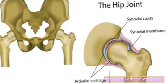
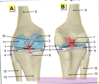
Category : Anatomy-Lexicon

External rotation

External rotation

External rotation is the rotation of a body part around its own axis and away from the body. The external rotation is the opposite movement to the internal rotation and can be carried out by means of ball joints. For example, when tightening the shoulders
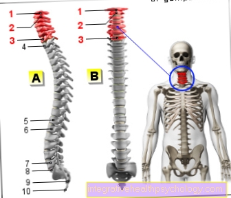
Hand nerves

Hand nerves

The hand nerves are responsible for the sensitive and motorized supply of the hand and arise from the arm nerve plexus (brachial plexus). A distinction is made between the median, radial and ulnar nerves. In the event of damage, there is usually a loss of function

Triangular disc

Triangular disc

- your orthopedic information portal. Here you will find information on the topic of the triangular disc explained to laymen.

Cervical vertebrae

Cervical vertebrae

The cervical spine is part of the spine and consists of seven cervical vertebrae. The first cervical vertebrae are called Atlas and Axis and are important for head movements and support the entire head. Often, accidents damage the cervical vertebrae, which is often the case

Lumbar spine (lumbar spine) x-ray with scoliosis

Lumbar spine (lumbar spine) x-ray with scoliosis

the medical information portal. Lumbar spine scoliosis x-rays.

Radial nerve

Radial nerve

The radial nerve is an important arm nerve that transmits information from the skin to the brain and is important for muscle movement. In the event of damage, the "hand drop" phenomenon occurs, in which the elbow joint is stretched

Rotator cuff

Rotator cuff

The rotator cuff is a group of muscles that surround the shoulder and thus stabilize the shoulder joint from all sides and allow the upper arm to move in a controlled manner in all directions.

Broad back muscle

Broad back muscle

The broad back muscle (M. latissimus dorsi) is a large, superficial back muscle that originates in the spine and attaches to the upper arm and sides of the ribs. Its main function is the adduction of the raised arm as well as the retroversion

Disc meniscus

Disc meniscus

A disc meniscus is a standard variant of the meniscus. Usually the menisci are shaped like a crescent moon. The disc meniscus, on the other hand, has a disc shape and is larger. Usually it does not cause pain and will

Trapezius

Trapezius

the sports medicine information portal. A lot of helpful information on the subject of cap muscles explained in an understandable way.

Cruciate ligament

Cruciate ligament

Humans have two cruciate ligaments in each knee joint, an anterior and a posterior cruciate ligament. They stabilize the knee joint and prevent excessive inward rotation. Cruciate ligament tears are one of the most common knee ligament injuries, although mostly

Quadriceps

Quadriceps

The quadriceps represent the largest and most powerful muscle in our body. The quadriceps is the most trained muscle in leg muscle training.The following are the functions, innervation, stretching and proper training of the quadriceps

Kneecap jumped out

Kneecap jumped out

The physician describes the condition of a popped kneecap as a dislocation of the kneecap or patella or a dislocation of the kneecap or patella. Such a dislocation of the kneecap is relatively common and has two basic mechanisms

Forefoot

Forefoot

The forefoot represents the forefoot part of the foot. It is formed by the big toe and the little toe. In the case of the little toes, a distinction is made between the base, middle and end joints. Joints known as interphalangeal joints can be found between the individual phalanges of the toes

Inner band knee

Inner band knee

The inner ligament of the knee joint connects the thigh to the shin. It is fused with the joint capsule and taut together with the outer ligament in the extended knee. In many sports, the inner band is due to strong pressure

Outer ligament of the knee

Outer ligament of the knee

The outer ligaments of the two knee joints connect the thigh to the fibula. Together, the inner and outer ligaments are also called "collateral ligaments". They provide lateral stability to the knee joint when it is extended and limit rotation

Taping the ankle

Taping the ankle

The ankle is one of the most commonly injured joints. These include bruises, slight stretching of the ligaments or also ligament tears and injuries to the joint capsule. Kinesio tapes gently massage the affected areas

Biceps brachii muscle / biceps muscle

Biceps brachii muscle / biceps muscle

The biceps muscle (muscle biceps brachii) called biceps for short, belongs to the flexor muscles and is located on the front of the upper arm. It is a two-jointed muscle that pulls over the shoulder and elbow joints. Frequent

Hindfoot

Hindfoot

Anatomically, the rear foot corresponds to a part of the tarsus. The term hindfoot comes from everyday clinical practice. Two of the tarsal bones are described here, namely the talus (ankle bone), the part of the ankle joint

Broken fibula

Broken fibula

In medicine, a fracture of the fibula is called a fibular fracture. Most often it occurs in connection with a fracture of the upper ankle joint. In the case of serious injuries, the connective tissue membrane between the shinbone can also be affected