Achilles heel
definition
The Achilles heel (tuber calcanei) forms the rear part of the heel bone (calcaneus). The name Achilles heel originally comes from Greek mythology. The calcaneus is the largest tarsal bone and, together with the talus, forms the rear foot. The Achilles heel serves as a protruding bone, the attachment point of the Achilles tendon.

anatomy
As a bony structure, the Achilles heel is a preferred attachment point for various muscles. The strong Achilles tendon, for example, attaches to the back of the bone. There is also a bursa at this point, the bursa tendinis calcanei (also bursa subachillea), which protects the tendon and bones from friction and damage. This bursa can sometimes become infected.
On the inner side of the Achilles heel one finds the processus medialis tuberis calcanei, a bone process which also serves as the muscle origin for the muscle flexor digitorum brevis and the muscle abductor hallucis. On the outer side there is also a bony process, the processus lateralis tuberis calcanei.
Various ligaments also attach to the lower side of the Achilles heel, such as the sole tendon plate (plantar fascia) and various joint stabilizing ligaments. Together with the ankle bone and the navicular bone, the heel bone forms the lower ankle joint.
Appointment with an expert in Achilles tendonitis?

I would be happy to advise you!
Who am I?
My name is I am a specialist in orthopedics and the founder of .
Various television programs and print media report regularly about my work. On HR television you can see me every 6 weeks live on "Hallo Hessen".
But now enough is indicated ;-)
Athletes (joggers, soccer players, etc.) are particularly often affected by the Achilles tendonitis disease. In many cases, the cause of the Achilles tendonitis cannot be identified at first. Therefore, the treatment requires a lot of experience. I focus on Achilles tendonitis.
The aim of every treatment is treatment without surgery with a complete recovery of performance.
Which therapy achieves the best results in the long term can only be determined after looking at all of the information (Examination, X-ray, ultrasound, MRI, etc.) be assessed.
You can find me in:
- - your orthopedic surgeon
14
Directly to the online appointment arrangement
Unfortunately, it is currently only possible to make an appointment with private health insurers. I hope for your understanding!
Further information about myself can be found at
function
The Achilles heel gives the back foot its characteristic shape. However, the main function is to be the insertion and origin of various muscles and ligaments. The Achilles tendon is located on the Achilles heel and is visible and palpable in slim people. The Achilles tendon connects the triceps surae muscle (calf muscle) to the rear foot.
When the calf muscle contracts, the tip of the toe tilts forward and the heel moves upwards (so-called plantar flexion). This enables walking on tiptoe, for example. The contraction of the calf muscles is one of the most important components of the walking process. In addition to plantar flexion, the triceps surae muscle also supports supination, the tilting of the foot to the inside.
On the inner side of the Achilles heel one finds the origin of the M. Flexor digitorum longum (long toe flexors) and M. abductor hallucis (big toe guide). The flexor digitorum longus muscle serves to bend the toes and the abductor hallucis muscle guides the big toe away from the other toes and also supports the function of the flexor digitorum longus muscle.
Various ligaments attach to the Achilles heel, such as the sole plate of the tendons, which connects the heel and the metatarsophalangeal joints. This connection stabilizes the longitudinal arch of the foot and thus enables stable walking and standing.
Diseases: Achilles heel
Achilles heel pain can have several causes. A broken Achilles heel or a tear in the bone, for example, can cause severe pain which is aggravated by heel strike. Muscle strains or overstretching are also painful, but usually go away after a short time.

Achilles heel pain can also arise due to a torn ligament or tendon and is also increased with movement. The collateral ligaments of the ankle and the Achilles tendon are particularly at risk. In addition to tears, tendinitis is also very painful and often requires treatment.
Inflammation can affect muscles as well as tendons, bones and bursa. Heel spur (also calcaneus spur) represents a special clinical picture. This is a new bone formation which, like a spur, can exert painful pressure on tendons and other structures.
Circulatory disorders in the context of arteriosclerosis, for example peripheral arterial occlusive diseases, can lead to pain when exercising due to a reduced blood flow. This is also called shop window sickness, as those affected often have to plan breaks when walking due to pain.
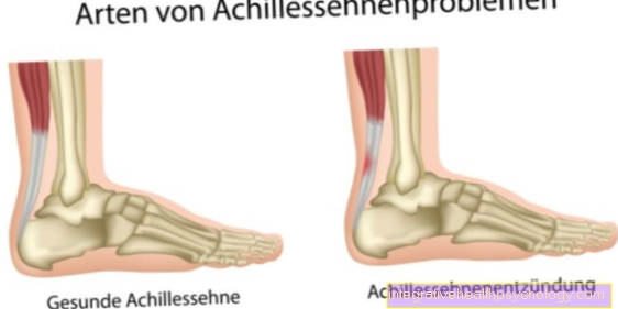
Diseases: inflammation of the Achilles heel
Inflammation in the Achilles heel can have different origins. A distinction is made between inflammation of the Achilles tendon (tendinitis) and inflammation of the Achilles tendon sheath (tendovaginitis).
Tendon infections often arise from (age-related) degeneration. The typical signs are pressure pain and stress pain. In this case, the pain arises when walking, and especially when standing on tiptoe. The diagnosis is usually made by means of an ultrasound examination or magnetic resonance tomography. Arthroscopy can also be performed if the findings are unclear.
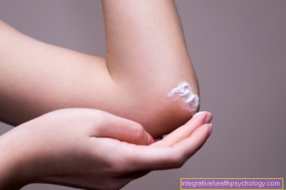
In mild cases, the therapy of choice consists of non-steroidal anti-inflammatory drugs and physiotherapy. In the case of severe courses or persistent inflammation, surgical measures can be considered.
Inflammation can also affect the bursa of the Achilles tendon (bursitis subachillae). Such inflammations often arise after trauma, recurring incorrect loads and infections. Such inflammations often make themselves felt as pain, swelling and / or overheating of the affected area.
If the inflammation was caused by bacteria, antibiotics may be used in addition to nonsteroidal anti-inflammatory drugs. To reduce the pain, the area can also be punctured to allow secretion to drain away.
Another form of inflammation is inflammation of the bones (osteomyelitis). This type of inflammation usually occurs after a bone fracture or an operation on the bone. In such cases, in addition to antibiotic therapy, the affected piece of bone usually has to be removed or replaced.
Diseases: posterior heel spur
The heel spur is a bone formation in the area of the heel. This new formation is often spur-shaped and can lead to discomfort while walking and pain. Such a new formation is observed especially in overweight people or people who (due to work) have to stand for a long time. A heel spur does not necessarily have to cause discomfort.
Only when the piece of bone affects the surrounding structures does it cause pain. Inflammation of the bursa tendinis calcanei (bursa between the Achilles heel and the Achilles tendon) is often incorrectly diagnosed as a calcaneal spur.
The easiest and fastest way to diagnose heel spur is an X-ray of the lateral foot. Various therapeutic approaches can be used for complaints. As a rule, a combination of pain relievers and adequate shoe inserts is preferred. In addition, physical therapy and cryotherapy can be prescribed. If the therapy is unsuccessful, one can try to destroy the heel spur with the help of shock waves or even consider surgical treatment.
Disease: Achilles heel strain
A strain of the Achilles heel is not possible per se, since it is a bone. However, the Achilles tendon can be strained in this area. In this case, the muscle is overstretched as a result of trauma or incorrect movement.
Tension of the Achilles tendon can occur preferentially during sporting activities with changing foot movements (such as soccer, rugby, tennis, etc.). Here the muscle is strained either too much or incorrectly. A muscle strain is characterized by pain when tensing or straining, but not when stretching the muscle.
If a muscle strain is suspected, the muscle should first be spared and cooled. In the further course, a (pressure) bandage and an elevated position can help.
In general, the PECH rule applies: break, ice, compression, elevation. If the symptoms get worse or have persisted for over a week without improvement, a doctor should be visited. Muscle strains can be avoided by warming up before exercise.
How can you tape the Achilles heel?
Taping the Achilles heel can be an effective relief in the context of certain sports or complaints.
There are two options for taping the Achilles heel. The first is mainly used to stabilize the ankle. You need two strips of tape for this.
The first strip is fixed from the middle of the sole of the foot to the back over the heel and further up on the back of the ankle. The portion from the heel of the foot to the ankle corresponds to the course of the Achilles tendon and should therefore be applied more tightly.
The second strip is cut in half and glued vertically over the first strip. The first horizontal stripe should be approximately at the level of the pain point on the heel, the second horizontal stripe just above it.
The second tape option is particularly suitable for calf pain. This requires three long strips of tape. The first strip is halved lengthways, except for a common end piece. This common end piece is glued to the sole of the foot in the area of the heel.
The remaining part (the two long thin strips) is taped around the calf muscle, which means that the calf is hugged by two strips. The next step is to stretch a long strip straight across your calf muscle to your heel. This strip runs from the hollow of the knee to the heel, where the first strip starts. Finally, the last strip of tape is divided into two. One half is glued vertically over the other strips at the level of the ankle. The second half is fixed to the sole of the foot in the area of the transverse arch (this corresponds to the base of the toes).
Additional information
Read more about orthopedics:
- Torn hamstring
- Knee pain - what do i have?
- bruise