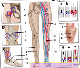
Vessels
Synonyms
- Latin: vas
- Greek: angio
- English: vascular wall
definition
A vessel in a body can be compared to a tube that transports the body fluids lymph and blood.
Depending on which liquid flows through this pipe system, a distinction is made between:
- Blood vessels and
- Lymphatic vessels.
All pipe systems in which other body fluids are transported are called “corridors” (lat. Ductus). This includes, for example, the tear duct, gland ducts, etc.

Blood vessel
The Blood vessel can be considered a flexible Imagine a tube in which the body's blood is transported. The individual blood vessels in humans close to form the complex blood circulation together.
The heart pumps oxygen- and nutrient-rich Blood over these into the periphery and from there it gets oxygen- such as low in nutrients Blood back to the heart.
Classification
The Blood vessels are divided into:
- aorta ( artery),
- Arteries (Arteries),
- Arterioles (small arteries),
- Capillaries (Hair vessels),
- Venules (small veins),
- Veins (Blood veins),
- upper / lower Vena cava (Vena cava superior / inferior)
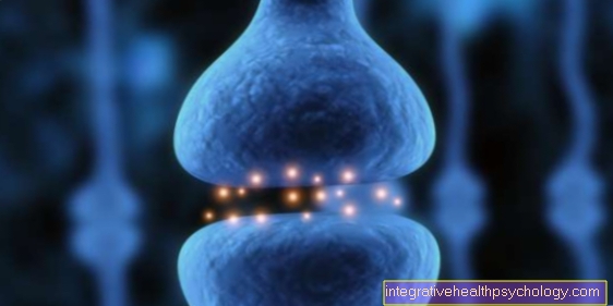
Structure of the vessels

The vessel wall in larger blood vessels is basically made up of three different layers:
- Tunica intima - intima
- Tunica media - Media
- Tunica externa or Tunica adventitia - Adventitia
Capillaries have a simpler structure. Pericytes, which are slightly changed contractile cells of the connective tissue, branch out around the thin endothelium. They also have the property of permeability, which other blood vessels do not have. This means that they are permeable to certain blood cells and molecules.
Intima: It is the inner layer of the vessel wall of the arteries, veins and lymph vessels. It consists of endothelial cells that are arranged in the longitudinal direction towards the vessel.
Their task is the exchange of gases, liquids and substances between the blood and the surrounding tissue. In addition, there is a subendothelial layer and a fenestrated or elastic layer (lat. Membrana elastica interna). Veins still have venous valves, which consist of two crescent-shaped leaflets that have their own connective tissue layer. The venous valves catch the blood flowing backwards and therefore ensure a continuous flow of blood to the heart.
The media: It is made up of smooth muscle cells, elastic fibers and collagen. Depending on the type of vessel, the tunica media has a more or less pronounced muscle layer, which is delimited inside and outside by a shell made of elastic connective tissue. Two types of arteries can now be distinguished:
- the elastic type arteries close to the heart, which are important for the Windkessel function and
- the arteries further from the heart of the muscular type.
The membrana elastica externa lies over the media as a delimitation to the adventitia. The veins are actually the same in the structure of the media. The only difference is the much thinner muscle layer.
The adventitia: it serves to embed the vessel in its surroundings and to stabilize it. For the most part it consists only of loose connective tissue, except in larger vessels it contains thin blood vessels, Latin Vasa vasorumwho are responsible for supplying the vessel wall. In the case of smaller blood vessels, this is not necessary as the supply takes place from the lumen of the vessel itself.
physiology
The blood vessels have the ability to enlarge or reduce the lumen of the vessel and thus the Bloodstream to modify. That's what they need Muscle layer the tunica media, which tense or relax the muscles through vegetatively supplied nerves.
This results in either:
- Vasodilation (Vasodilation) or one
- Vasoconstriction (Vasoconstriction).
Since the arteries have a much thicker muscular layer, this phenomenon applies more to them and less to veins. Using this simple mechanism, the body can control the available blood volume, contribute to temperature regulation or improve the oxygen supply in the tissue.
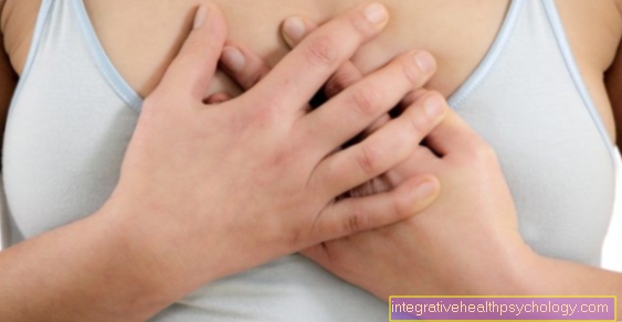
In the blood vessels there is a physiological one Blood pressure, which in the arterial vascular system between 80 and 120 mm Hg and does not exceed 10 mm Hg in the venous system.
clinic
There are many Diseasesaffecting the vascular system.
This includes, for example:
- arteriosclerosis,
- Occlusive diseases,
- inflammatory vascular disease (Vasculitis),
- functional Circulatory disorders (Agrocyanosis, Raynaud's Syndrome, Erythromegaly),
- Varicose veins,
- Thrombosis;
Neovascularization
All forms of Neoplasm of blood vessels in the adult organism are so called. This includes:
- angiogenesis,
- the vasculogenesis and
- the arteriogenesis.
In angiogenesis arise through Sprout- or Splitting processes new blood vessels from already formed. She plays a vital role in Wound healing. Vasculogenesis is important in the embryonic period. Here vascular structures develop through circulating Stem cells, so called angioblasts, which continue to mature into endothelial cells. Arteriogenesis is the formation of arteries and small arterioles. By recruiting smooth muscle cells, a complete vessel wall is created. The formation of new veins takes place in a similar manner.
Lymphatic vessels
Lymphatic vessels are very similar to blood vessels. However, they do not transport blood, but lymph, this is a liquid that is located in the tissue and contains small amounts of protein. In the conduction system of the lymph are Filter stations, so-called Lymph nodes, interposed.
construction
There are four types of vessels:
- The Lymph capillaries represent the smallest unit in Lymphatic system They have their beginning in the intercellular space (interstitium). they consist of Endothelial cellsthat overlap like roof tiles. As a result, they form a lumen approx. 50 µm in size. Anchor filaments fix the lymph capillaries in the surrounding tissue and also keep the lumen of the vessels open. In the lymph capillaries the Lymph formation instead of. This is created by absorbing the tissue fluid in the cell space.
- The Precollectors are the next larger lymph vessels that arise from the union of several lymph capillaries. The precollectors transport the lymph to the collectors with the help of isolated muscle cells. They are also involved in lymph formation, as they also absorb tissue fluid.
- Several pre-collectors combine to form one collector. The collectors are solely responsible for transporting the lymph from the existing lymph vessels. In anatomy, they are very similar to veins with a three-layer wall structure and valves. The valves prevent the lymph from flowing back and thus ensure a centrally directed lymph flow. The area between two valves is called the lymphangion ("lymph heart"). At rest, this contracts every 10–12x / minute, which pushes the lymph into the next section. Furthermore, the collectors are divided into superficial and deep collectors. The superficial collectors are located in the subcutaneous fatty tissue. They absorb the lymph from the skin and subcutaneous tissue. The deep collectors are located within the fasciae in the extremities and the trunk wall. They transport the lymph from muscles, ligaments, joints and bones. Gut collectors, as the name suggests, collect the lymph from the intestines.
- The Lymphatic collecting strains are the largest lymph vessels in the body. They are divided into lymphatic strains of the upper and lower halves of the body. The tracheal trunk and the thoracic duct are among the lymphatic trunks. They absorb the lymph from the collectors. Their final stretch is the vein angle near the heart, where they flow into the venous blood circulation.
Lymph vessels of the same level, for example superficial collectors in the subcutaneous fatty tissue, are interconnected by so-called Anastomoses connected. Such vessels, which are located in different levels, such as superficial and deep collectors, establish a connection with one another through so-called Perforation vessels here. This creates an exchange of fluid flowing from the deep to the superficial lymph vessels. In the Lymphatic drainage by means of massages, this property is made use of. Anastomoses are especially important to avoid lymphedema. They serve as diversions if there is a congestion in a system or the lymphatic transport is completely interrupted.
task
The Lymphatic system is responsible for collecting the protein molecules and the fluid remaining in the surrounding tissue and transporting them to the venous line system. Additionally it is for that Fat digestion necessary.
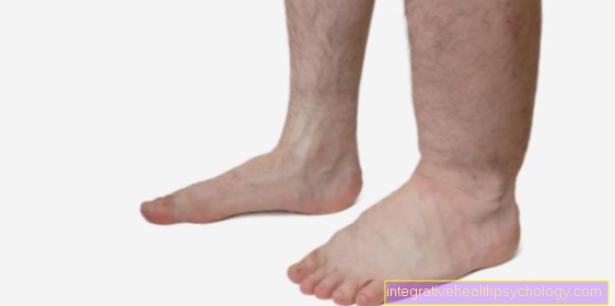
In this process, the majority of the fats ingested from food are packed by cells in the small intestine in so-called chylomicrons and then transported into the blood via the lymph vessels. If there is a backlog in the lymphatic system, for example by a Right heart failure, this can lead to lymphedema especially in the legs.
As already mentioned, the lymph is important for protein transport. If the protein remained in the tissue, the colloid-osmotic pressure in the surrounding tissue (the interstitium) would change and blood cells could also get into the interstitium. This would have one Lack of volume (Hypovolemia), which in the worst case can be life-threatening State of shock can trigger.


.jpg)