Aneurysmal bone cyst
definition
The aneurysmal bone cyst belongs to the category of benign bone tumors. It is a blood-filled cyst located in the bone, which is divided into several individual cavities by septa, that is, chambered.
An aneurysmal bone cyst usually occurs between 10 and 20 years of age, so it is a bone lesion in young people. Much of the aneurysmal bone cyst is definitely diagnosed before the age of 20. Both sexes are equally affected.

An aneurysmal bone cyst can generally form on any bone in the human body. Predisposed areas are, however, the thighbone (lat. Femur) and one of the two lower leg bones, namely the shin (lat. Tibia). In each of these two bones, the area of the metaphysis, i.e. the part between the bone shaft and the joint-forming bone portion, is the most common place of manifestation. In addition, an aneurysmal bone cyst often appears in the spine.
The most important differential diagnosis to aneurysmal bone cyst is the juvenile bone cyst. However, a clear differentiation is possible with the help of imaging procedures.
Read more on the subject here Juvenile bone cyst
causes
The causes of an aneurysmal bone cyst can be defined relatively clearly. There is a variant that applies in about 80% and which is a primary idiopathic bone cyst. Idiopathic means that the cause of the cyst formation is not known or cannot be defined.
The second possibility for the cause of an aneurysmal bone cyst is that it was secondary to other malignant bone lesions or was a concomitant phenomenon of other diseases. In addition, there are no significant risk factors or gender predispositions in connection with the aneurysmal bone cyst that could have a causal influence.
Symptoms
The aneurysmal bone cyst is a relatively uncomfortable bone lesion. There is seldom pain and swelling. In exceptional cases, however, swelling can be so pronounced that it can simulate a tumor, i.e. a mass that is visible from the outside. Often those affected do not even notice that they have an aneurysmal bone cyst. It is often only recognized when the affected bone breaks as a consequence or accompanying symptoms. The cyst makes the bone less stable and resistant, so that it can break with disproportionately little stress. The aneurysmal bone cyst is therefore often an incidental or incidental finding when taking X-ray or MRI images due to other indications.
Since the aneurysmal bone cyst is a benign bone tumor, there is no weight loss, night sweats or fever, as would be expected with a malignant tumor.
If you would like to know more about the differentiation from malignant bone tumors, read more on the subject here Bone cancer
diagnosis
The diagnosis of an aneurysmal bone cyst is made using imaging techniques.
A clinical diagnosis is difficult or impossible, as there are no typical symptoms that would be sufficient for a clinical diagnosis alone. In addition, the aneurysmal bone cyst manifests itself very variably in those affected. However, if there are indications in the form of pain or swelling on predisposed bones or if there is a fracture that was probably provoked by a bone cyst, an X-ray image is first made in 2 planes. Here you can clearly see the bony lesion, which is mostly located in the area of the metaphysis and can be clearly defined. One also speaks of an “osteolytic” lesion, i.e. a breakdown or dissolution of the bony structure in the area of the cyst.
If the findings after the X-ray are still not entirely clear or if the findings cannot be differentiated from the differential diagnosis of a juvenile bone cyst, an MRI image is taken. The MRI shows a blood-filled bone lesion which, in contrast to the juvenile bone cyst, is characteristically chambered, i.e. separated by a septum. A diagnosis can ultimately also be confirmed by an open biopsy.
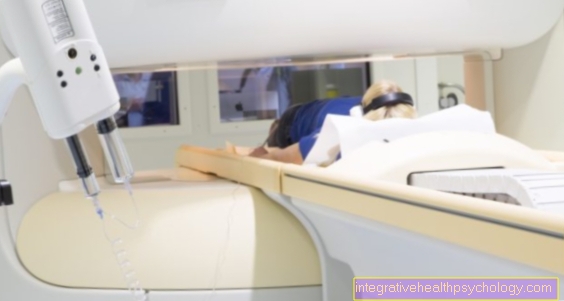
MRI
As part of the diagnosis of an aneurysmal bone lesion, an MRI image is only made after an X-ray has been taken beforehand. Characteristically, the aneurysmal bone cyst presents itself in the MRI as a bony, blood-filled lesion which is chambered by septa. It is found on long bones, such as the thigh, mostly in the area of the metaphysis. A typical phenomenon of the aneurysmal bone cyst in the MRI is called “fluid-fluid-level”. This describes a so-called stratification phenomenon, which is caused by the sinking of components in the blood that is located in the cyst. The stratification phenomenon can look like a further subdivision of the already chambered cyst, since the deposited blood components or sediments are represented as lines. The differential diagnosis of a juvenile bone cyst due to the existing septation can be ruled out very well in the MRI image, since the chambered bone cyst is a feature of the aneurysmal bone cyst.
treatment
The only conservative treatment approach that remains is symptom-oriented pain therapy if necessary. Which pain reliever is most suitable for you depends, among other things, on previous illnesses or allergies. Therefore, discuss the pain therapy with your doctor.
Instead, the aneurysmal bone cyst has to be treated surgically. Surgical treatment of the aneurysmal bone cyst involves clearing out the blood-filled cyst. The removal of the bone cyst is usually accompanied by careful scraping, known as curettage in technical terms. In addition, the lesion is filled with so-called cancellous bone, a material that is physiologically located in the interior of the bone.
As an alternative to cancellous bone material, the cyst can also be initially filled with bone cement. In a second operation, the cement can be replaced at a later point in time with the body's own cancellous bone, for example from the iliac crest.
One possible therapy method that is rarely used is irradiation of the cyst. Since those affected are usually very young, this approach has no significant benefit due to the high radiation exposure and is therefore only used in exceptional cases with aggressive cyst forms. The treatment of an aneurysmal bone cyst is generally relatively difficult because few bone cysts respond well to therapy and a few years later there is rarely no recurrence. Since most aneurysmal bone cysts are also primarily idiopathic, i.e. of unknown cause, no direct causal therapy is possible.
When is an operation necessary?
Surgical treatment of an aneurysmal bone cyst is almost always indicated. Surgery is only not necessary if there are no complaints, no susceptibility to fractures and a spontaneous regression tendency. Since this is almost never the case, conservative treatment in the form of pain therapy and waiting to see whether the cyst regresses is not enough. Surgical care is not always successful in the long term due to the high rate of recurrence, but it is the only way to combat the aneurysmal bone cyst for the time being. As soon as an aneurysmal bone cyst has been diagnosed as the main or secondary finding in the X-ray and / or MRI, an operative procedure can be planned individually.
Duration
The time it takes for an aneurysmal bone cyst to heal varies. It depends on how pronounced the findings are, how old the person concerned is and whether the bone involved has already broken, which is related to the aneurysmal bone cyst. The duration of therapy, including the healing process, usually extends from weeks to months. Precise information is rather difficult to give, as the therapy method is variable and those affected respond differently to it. It should also be said that the aneurysmal bone cyst can reappear even after it has healed completely. Children who developed the aneurysmal bone cyst before the age of 10 are particularly prone to local recurrence. Malignant degenerations that would take longer to heal and, above all, take longer to treat, only occur relatively rarely in the context of an aneurysmal bone cyst.
Localization of the bone cyst
Bone cyst in the jaw
The jaw as a place of manifestation of the aneurysmal bone cyst is rather rare. Typical locations are instead the thighbones (lat. Femur), the shin (lat. Tibia) and the spine. In less than 2% of cases, however, an aneurysmal bone cyst occurs in the jaw. The cyst forms more often in the lower than in the upper jaw. The symptoms of aneurysmal bone cyst in the jaw range from symptom-free cysts to disfiguring deformities of the face due to the enormous growth of the cyst.
In the jaw, it is particularly important to rule out the possibility of a vascular cyst. Once this has been done, all diagnostic measures for an aneurysmal bone cyst of any location apply: imaging in the form of X-rays and MRI, as well as a biopsy for further examination of the tissue. Accordingly, general treatment in the form of surgical treatment for an aneurysmal bone cyst in the jaw is necessary.
Bone cyst in the thigh
The aneurysmal bone cyst in the thigh is considered a typical localization. Sometimes a cyst in the thigh becomes noticeable due to pain, which can radiate into the legs and back. Otherwise, an aneurysmal bone cyst on the thigh can also be discovered as an incidental finding.
In young people up to the age of 20, aneurysmal bone cyst in the thigh tends to have fewer symptoms. Older people have a significantly higher risk of fractures. It is not uncommon for them to present themselves with a fracture of the thigh that has arisen due to an aneurysmal bone cyst. Due to the cystic mass, the bony structure is weakened, less stable and prone to breakage when the load is even lower. The aneurysmal bone cyst on the thigh is diagnosed with an X-ray and MRI image and then treated surgically.