Air in the abdomen
What is air in the abdomen?
Free air in the abdomen (med. Peritoneal cavity) is also known as pneumoperitoneum. A pneumoperitoneum can be created artificially by a doctor, for example as part of an operation, and in this case is referred to as a pseudopneumoperitoneum. But pathological processes or injuries to the abdominal cavity can also lead to this clinical picture.

The reasons
Normally the air in the abdomen is only in hollow organs such as the intestines or the urinary bladder. Air outside of hollow organs does not occur in healthy people. Doctors then refer to this air as "free air".
A pneumoperitoneum can also be created artificially by a doctor. This occurs in minimally invasive procedures such as laparoscopy. Here, the surgeon pumps up the abdomen with a gas in order to create a better view and more space during an operation. This air can remain in the patient's abdomen for a few days and has no disease value.
The cause of free air in the abdomen is a perforation or an injury to a hollow organ. An example of this is the perforation of a gastric ulcer or the perforation of an inflamed appendix. Diverticulitis is another high risk of perforation of a hollow organ. This is an inflammatory protrusion of the large intestine. Older patients in particular are affected by this. If a perforation occurs, those affected suffer from severe abdominal pain and the abdominal wall is as hard as a board (so-called acute abdomen).
However, a perforation can also arise from an invasively growing tumor. Free air can also accumulate in the abdomen if the outer covering of the abdomen is damaged and air can penetrate the body from outside.
Read our topic: Tumor in the abdomen
After an operation
During an operation in the abdomen, the abdominal cavity is opened and the surgical procedure is carried out. This procedure is also called a laparotomy. After sewing and closing the abdominal wall, there may be free air in the abdomen.
A common cause of air in the abdominal cavity is a laparoscopy or laparoscopy. Nowadays, more and more interventions are performed using minimally invasive methods. This means that you only make small incisions during the operation so that the body can recover more quickly afterwards. At the beginning of the laparoscopy, three to five liters of carbon dioxide are pumped into the patient's abdominal cavity using a special machine. For this, the patient's abdomen is punctured with a needle and the gas is introduced through it. As a result, the patient inflates, the abdominal wall rises and the organs separate from each other. As a result, the surgeons have a better overview of the abdominal organs and enough space to operate. At the end of the operation, the gas is pumped out, but not all of the carbon dioxide can be removed and some remains as free air in the abdomen. This air can stay there for up to two weeks before it is gradually absorbed through the intestinal wall and finally exhaled by the patient. Typically, after the procedure, patients feel bloated and feel pressure in their abdomen.
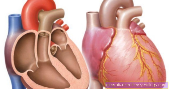
In general, carbon dioxide is considered a suitable gas and has prevailed over helium and nitrous oxide in surgery. In rare cases, however, the creation of the pneumoperitoneum can also result in complications. The introduced gas exerts a certain pressure in the abdomen, which compresses the large venous blood vessels and can disrupt the return flow of blood to the heart. As a result, the heart function can be restricted. Therefore this method is unsuitable for people with heart disease.
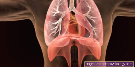
Even patients with impaired lung function (asthma or chronic obstructive pulmonary diseases) cannot undergo minimally invasive surgery because they cannot adequately breathe the remaining CO2.
Read more on the subject here Laparoscopy.
After a caesarean section
During a caesarean section, the abdomen is surgically opened and the child is taken out of the uterus.
As with all abdominal operations, air enters the abdomen, which accumulates and can still be detected a few days after the operation.
However, this is completely normal and does not need any further treatment, but it often makes women feel bloated and suffer from abdominal pain.
Learn more about the topic: Pain after a caesarean section.
The symptoms
Free air in the abdomen increases the pressure and thus leads to discomfort. Symptoms depend primarily on the amount of free air and the cause.
Free air that remains in the abdominal cavity after an operation usually causes only minor discomfort. Patients feel bloated and feel uncomfortable pressure in their abdomen.
The perforation of an abdominal organ, on the other hand, suddenly causes severe abdominal pain. In addition, the abdominal wall is clearly hardened. In addition, those affected are in a generally poor condition, which can develop into circulatory shock. The pain can sometimes be so severe that those affected feel sick and vomit.
Doctors refer to this complex of symptoms as acute abdomen (lat. belly). An acute abdomen is an absolute emergency and requires immediate medical attention.
If the small or large intestine is perforated, intestinal contents enter the abdominal cavity, causing peritonitis (Latin peritonitis) arises. Patients develop a high fever, nausea, vomiting, and constipation or diarrhea.
more on the subject Acute abdomen can be read here.
Pain
Small amounts of free air, such as that left in the abdomen after an operation, cause no or only slight pain. However, if free air gets into the abdominal cavity through the perforation of a hollow organ, those affected feel very severe abdominal pain that occurs suddenly.
The pain is described as burning and dull and very difficult to localize. The patient moves into a relieving position and tries to alleviate this pain somewhat by bending.
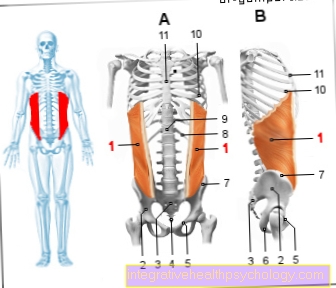
When examined by a doctor, the hard abdominal wall is particularly noticeable, which, along with severe pain, is one of the main symptoms of an acute abdomen.
The diagnosis
Doctors can use imaging tests to diagnose pneumoperitoneum. The air in the abdominal cavity can be easily visualized in an X-ray or a computer tomography (CT) scan of the abdominal cavity and even very small amounts can be easily detected.
In an X-ray that was taken while standing, you can see the open air as a narrow sickle under the diaphragm. Depending on the patient's position, the free air can be detected in the CT as an air bubble under the diaphragm (standing image) or as a lateral lightening over the liver (left lateral position).
treatment
If the free air in the abdominal cavity is due to a recent surgical procedure, no treatment is required. The gas is absorbed through the intestinal wall, gets into the blood and is exhaled through the lungs.
In the case of a pathological pneumoperitoneum, therapy is based on the cause.
If the air gets into the abdominal cavity through injury to the peritoneum, the wound is closed and treated. Tumors that invasively grow into organs and ultimately damage them in such a way that perforation occurs are removed surgically if possible. Then an attempt must be made to sew the perforated organ back together.
The perforation of an abdominal organ can also be caused by injuries or inflammatory processes in the body (e.g. diverticulitis, stomach ulcer).
A perforation is always considered an absolute emergency and must be operated on immediately. During the operation, an attempt is made to sew up the hole. This is followed by drug therapy with antibiotics to treat or prevent peritonitis.
Prognosis
The prognosis of pneumoperitoneum depends on the cause. Free air that has entered the abdominal cavity through an operation is harmless and disappears after a few days without treatment.
If a perforation of an abdominal organ leads to free air in the peritoneum, action must be taken quickly, as this is a potentially life-threatening condition.
If peritonitis has already developed, life-threatening blood poisoning up to multi-organ failure can occur.