Diagnosis of back pain
introduction
Since back pain can have a wide variety of causes, it is very important to carry out a detailed diagnosis in order to identify the underlying problem of back pain in order to then be able to treat it successfully.
This diagnosis of back pain includes a thorough anamnesis (interview) as well as a physical examination and possibly technical procedures.

History of back pain
Since there are different causes of back pain, the anamnesis is particularly important. Many back pain has psychological reasons (please refer: Back pain and psyche) or are at least aggravated by psychological complaints, so above all value should be placed on a social history.
The occupational history also plays an important role in the diagnosis of back pain, in order to determine whether the patient is exposed to heavy loads in his job, for example whether he has to remain in a standing or sitting position for a long time or lift heavy objects.
In addition, when making a diagnosis, it is important to precisely define the back pain, precisely because there are so many different forms.
It is important for the doctor to ask
- when the back pain occurs,
- how often,
- where exactly,
- whether the pain radiates to other parts of the body,
- how bad the pain is
- whether they improve or worsen in certain situations,
- how long they last, since when they have existed,
- whether there are any further complaints.
It is of great help if a person concerned keeps a so-called "pain diary" before his medical diagnosis, in which he lists all these points when the back pain plagues him. As a result, a lot can often be excluded and the diseases that are still possible can then be tested more specifically.
Also read the article on the topic: Psychosomatic back pain
Physical examination for back pain
This medical history is usually followed by a physical exam.
This includes checking the muscle strength of the back muscles and abdominal muscles and the mobility of the back in the various body axes. In addition, it should be checked whether the patient has local tenderness. Differences in leg length are also apparent in the clinical diagnosis. A neurological examination (examining the functionality of the nerves that emerge from the spine) can provide information as to whether the spine is damaged and, if so, in which area.
Imaging for back pain
As a rule, these methods are sufficient to find out the reason for the back pain. In some cases, however, more extensive diagnostics are necessary. The various imaging methods are available for this purpose.
First of all, the standard is the X-ray. This is less stressful for the patient and already gives the doctor a good insight into whether there are any abnormalities in the spinal column. The CT and MRI are more complex, but also more informative.
With these measures, either with or without contrast medium, sectional images of the chest (thorax) so that you can assess bony structures as well as soft tissues and nerves.
A contrast medium should only be used if there is reasonable suspicion of an inflammation or a tumor, as it exposes the patient's organism to additional stress and quite a few people are allergic to contrast medium. Another area in which contrast media can be used is what is known as myelography, which is carried out when a pathological process within the spinal cord itself is assumed.
This is where the contrast agent is injected into the area where the nerves leave the spinal canal. Sometimes a blood test can also be useful, as this can use certain parameters to provide information about whether there is an inflammation or a tumor in the body.
However, one should be careful not to start too lightly with a pronounced apparatus-based diagnosis. Many people have findings that differ significantly from the norm, but are by no means the cause of back pain. However, if this is misinterpreted as a reason, a long, stressful therapy can follow, which is not only unnecessary, but also does not bring about any improvement. Since one has “rushed” to this first result, the actual cause of the pain often remains undetected.
In such cases, this is often psychological or simply due to poor posture and the resulting tension and these problems are then not even addressed.
Read more about the topic here: Exercises for back pain
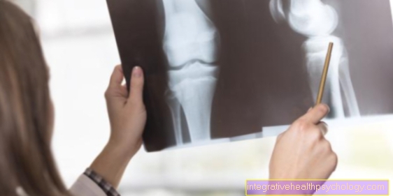
X-ray to diagnose back pain
For back pain, an x-ray can be helpful if the cause is suspected to be in the bones.
If, for example, scoliosis, i.e. a faulty curvature of the spine, is the cause, a diagnosis with an X-ray is always required. This is important to determine the extent of the scoliosis.
Also for so-called function recordings, e.g. X-rays are well suited for images in a position of bending back and forth.
The more common causes, such as Herniated discs, an x-ray is not necessary. It should never be carried out without a sufficient reason, i.e. without a correct indication, as the radiation exposure should not be underestimated.
You might also be interested in: Scoliosis
Appointment with a back specialist?

I would be happy to advise you!
Who am I?
My name is I am a specialist in orthopedics and the founder of .
Various television programs and print media report regularly about my work. On HR television you can see me every 6 weeks live on "Hallo Hessen".
But now enough is indicated ;-)
The spine is difficult to treat. On the one hand it is exposed to high mechanical loads, on the other hand it has great mobility.
The treatment of the spine (e.g. herniated disc, facet syndrome, foramen stenosis, etc.) therefore requires a lot of experience.
I focus on a wide variety of diseases of the spine.
The aim of any treatment is treatment without surgery.
Which therapy achieves the best results in the long term can only be determined after looking at all of the information (Examination, X-ray, ultrasound, MRI, etc.) be assessed.
You can find me in:
- - your orthopedic surgeon
14
Directly to the online appointment arrangement
Unfortunately, it is currently only possible to make an appointment with private health insurers. I hope for your understanding!
Further information about myself can be found at
MRI of the spine
Magnetic resonance imaging, i.e. an MRI, is a very helpful method when it comes to assessing possible so-called soft tissue damage on the back. The most common of these is the herniated disc, for which an MRI is the best option for detection. The MRI shows muscles and ligaments in particular very well.
As a rule, only parts of the spine are examined, depending on where the pain is localized. Lie as e.g. Lower back pain with suspected herniated disc (this is most common in this region), an MRI scan of the lumbar spine is ordered.
Before an MRI scan, it should be carefully considered whether this is necessary. Due to the long duration of the exposure and the exposure to noise, it is an uncomfortable situation for the person to be examined. Therefore, the guideline is that an MRI exposure should only be carried out if the back pain is still present after about 6 weeks and none Cause was found.
Of course, there are exceptions to this, which should be discussed with a doctor. An MRI should be done earlier, especially if there are signs that suggest an entrapment, such as long-term numbness in one leg.
also read: Magnetic resonance imaging
CT of the spine
Computed tomography, i.e. CT, of the spine is often done in the case of back pain when a fracture of one or more vertebral bodies is suspected. In the case of a herniated disc, a so-called follow-up is often required to check whether the tissue recovers after a certain period of time, usually under therapy. A CT is also usually used for this, since this type of imaging is much faster and less complex. Most types of back pain do not require a contrast agent on a CT image.
You can also find out more at: Computed Tomography
Myelography for back pain
Myelography is a procedure in which contrast agent is first injected into the spinal canal and then an X-ray image of the spine is performed.
This enables the spinal cord and, above all, the surrounding envelope, the so-called spinal canal, to be displayed particularly well. Nowadays, with the availability of MRI and CT imaging, myelography has decreased.
However, if the back pain is caused by a blockage of the spinal canal, for example, myelography can be helpful for precise localization and size estimation. In most cases, this imaging is carried out accordingly after another recording that did not provide sufficient information.
Read on below: Myelography
Discography for back pain
In a discography, contrast agent is injected into an intervertebral disc in order to then observe how it is spreading in X-rays. The person to be examined is lightly anesthetized. Back pain is often caused by intervertebral discs, i.e. protrusions or displacements. This type of pain is usually caused by pressure from the intervertebral disc on the nerves emerging from the spinal cord. Discography is an invasive type of diagnosis and therefore only to be carried out if it is clearly necessary.
Recommendations from the editorial team
More information about back pain:
- Back pain
- Back pain
- Causes of back pain
- Therapy of back pain
- Lumbar spine pain
- Lower back pain
- Lower back pain
- Back pain upper back
- Back pain middle back
- Symptoms of back pain
- Back pain in pregnancy
- Back pain what to do?
- Torn hamstring back