Cardiovascular system
Synonyms
Blood circulation, large body circulation, small body circulation
Medical: Cardio-pulmonary circulation
English: cardiovascular system
Also read: Circulatory weakness
definition
The cardiovascular system can be imagined as a combination of two individual sections (the small and large body circulation) that are connected in series.
They are connected through the heart. The great circulation supplies the body with nutrients and goes from the left side of the heart to the mouth in the right atrium. The small circuit goes from the right heart through the lungs for gas exchange and flows into the left atrium.

Illustration of the cardiovascular system

- Superior vena cava -
Superior vena cava - Lower vena cava -
Inferior vena cava - Ascending aorta -
Pars ascendensaortae - Aortic arch -
Arcus aortae - Pulmonary artery trunk -
Pulmonary trunk - Left pulmonary artery -
A. pulmonalis sinistra - Right pulmonary veins -
Vv. Pulmonary dextrae - Left pulmonary veins -
Vv. Pulmonary sinastrae - Mitral valve - Valva mitralis
- Aortic valve - Valva aortae
- Pulmonary valve -
Valva trunci pulmonalis - Right atrial ventricular valve
(Tricuspid valve) -
Tricuspid valva
Great Cardiovascular System - (red)
Small cardiovascular system - (blue)
You can find an overview of all Dr-Gumpert images at: medical illustrations
Structure of the cardiovascular system
The cardiovascular system consists roughly of the blood vessels and the heart as a muscle pump (Task of the heart), which allows blood to circulate around the body and provide oxygen and nutrients to tissues. The Organs and body tissues consume oxygen. Accordingly, new, oxygen-rich blood has to be supplied constantly. This will be the "Used" blood through the veins back to the heart transported. The many smaller veins from the extremities and organs unite in the abdomen and in the upper chest in the large vena cava (Superior vena cava and inferior). This opens into the from above and below right atrium of the heart. From there, the blood enters the right ventricle through a heart valve and is then passed through a another heart valve in the ejected right and left lungs. There the blood is again enriched with oxygen. The blood then travels from the lungs to the left atrium of the heart, through a valve into the left ventricle and then through the major artery (aorta) back into the big cycle. From there, it is distributed throughout the body via the arteries and supplies oxygen and nutrients to all organs and extremities.
Depending on Environmental conditions (Heat, cold, exertion, rest) the heart changes its beating rate. The blood vessels can expand expand or pull together. When it is cold outside, the blood vessels in the extremities contract so that less blood flows into them and the body does not cool down as quickly (centralization). In contrast, when the heat is on, the vessels widen because the body tries to give off the excess heat and the Keep core body temperature constant. Sweating also serves this purpose. During physical exertion, the vessels, especially the vessels in the muscles, also widen because they requires more oxygen during exertion. Accordingly, the blood volume is distributed over one larger cross-sectional area. The heart must now beat faster in order to allow sufficient volume to circulate in the vascular system. In athletes, exercise increases the heart rate over time. This allows it to eject more volume per stroke, so that it needs a lower stroke frequency both at rest and during exercise. This often explains this considerably lower resting heart rate of athletes. Overall, the cardiovascular system is very complex and consists of the smallest vessels (Capillaries) to the large arteries and veins that carry blood to and from the heart. The regulation of the cardiovascular system is also very complex and, in healthy people, can adapt very flexibly to different conditions.
Important details about the cardiovascular system
Arteries are called the vessels that lead away from the heart,
Veins are vessels that flow to the heart.
These expressions say Nothing about the oxygen content!
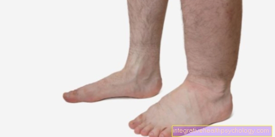
Are the veins - especially the superficial ones of the leg - no longer able to transport the blood back to the heart quickly enough, then arise Varicose veins (Varices).
Slowing blood flow in a deep vein can cause a Blood clot (thrombus) form the clinical picture of thrombosis evokes.
If such a blood clot loosens and becomes with the blood circulation in the lung worn then can be life-threatening Pulmonary embolism arise.
Classification of the vessels in the cardiovascular system
The vessels are divided into the following structures:
- Arteries (elastic type, muscular type)
- Arterioles (small arteries)
- Capillaries (vessels with the smallest diameter)
- Venules (small veins)
- Veins (small, medium and large veins; capacitance vessels)
These structures merge continuously.
The information in brackets after the terms will be explained in more detail later.
General wall construction of blood vessels:
In principle, the wall of arteries and veins consists of three layers:
- Tunica externa (outer layer)
- Tunica media (middle layer)
- Tunica intima (inner layer)
The outer layer or connective tissue layer contains nerves as well as some small (for the vessel itself) supplying blood vessels (Vasa vasorum). The middle layer consists mainly of changing parts. There are smooth muscle cells, elastic fibers and collagen fibers. The inner layer consists of a single-layer, flat cell structure.
In some arteries and veins, a so-called internal elastic membrane separates these two structures. Exceptions to these common features are capillaries and venules. These only have a single-layer wall. The only differences between arteries and veins are the properties of the wall layers. Arteries have a pronounced internal elastic membrane in their inner layer (Tunica intima), But not veins. The middle layer (Tunica media) is well developed in arteries. This structure is rather weak in veins. The outer layer (Tunica externa) is sparsely developed in arteries in contrast to the veins.
Arteries
Arteries per se are divided into an elastic type and a muscular type. Elastic-type arteries are usually strong arteries close to the heart that consist primarily of elastic fibers. These types of arteries represent an important factor for a continuous blood flow. They achieve this through the so-called wind chamber function. On the other hand, arteries of the muscular type are arteries distant from the heart, which regulate the blood flow to the organs by changing the diameter of the vessels.
Read more on the topic: artery
Arterioles
Arterioles are small arteries whose middle layer consists of a maximum of 2 layers of (smooth) muscle cells. They have an influence on the vascular resistance, especially in regions remote from the heart, and thus have an important influence on blood pressure.
Capillaries
Capillaries have the smallest diameter of all blood vessels. This is around 5-10 µm. This is of crucial importance because the diameter of a red blood cell (erythrocyte) is approx. 7.5 µm and the lumen is therefore just big enough for the erythrocytes to flow through. The capillaries run through the body like a network. So they are able to ensure the supply of all body cells. The capillary network is particularly pronounced in the lungs, kidneys and organs with hormonal functions, since the metabolic activity is particularly high here. The wall of the capillaries consists of a layer of flat endothelial cells that line the interior of blood vessels.
Read more on the topic: Capillaries
Venules
Venules, i.e. small veins, initially have roughly the same (wall) structure as capillaries. Their diameter is 15-500 µm. As a result, an exchange of substances is still possible in this section. This is why one speaks of postcapillary venules in this context. The wall construction just mentioned, however, can change progressively. For example, collecting venules have the familiar three-layer wall structure. Venules and arterioles are the smallest blood vessels that can still be seen with the eye.
Veins
As mentioned above with the classification of the vascular system, a distinction is made between small, medium-sized and large veins. Large veins can reach a diameter of up to 10 mm. Their main task is to transport the blood back to the heart. Arteries that carry blood away from the heart usually run parallel to veins and are roughly the same size. The wall of the veins is each much more elastic and thinner. This has the consequence that the inner radius of these vessels is also significantly larger. The fact that veins have such a thin wall is also due to the fact that one speaks here of a low-pressure system. The physical pressure load in veins is much lower than in arteries. They also make it difficult to distinguish the structures of the tunica intima, media and externa in the venous system. An additional specialty of veins are their valves.
Read more on the topic: veins
Venous valves are found in small and medium-sized veins. They are primarily responsible for ensuring that blood flow back to the heart is ensured. Venous valves themselves consist of a kind of “bulge” of the tunica intima, the innermost layer. Their functionality is similar to a valve. This opens the valves for blood to flow back to the heart. Blood flowing away from the heart causes the valves to fill and occlude.
Improve the cardiovascular system
To train your own cardiovascular system, a Cardio exercise which one from Endurance sports consists. This should Training units of at least 30 minutes to get voted. Sports that are suitable for cardiovascular training include jogging and swimming, as well as training sessions on the Treadmill, exercise bike, cross trainer or Stepper. Also Rowing, cross-country skiing or Nordic walking are possible. It is important that the training takes place regularly.
Effects
Cardiovascular training has many positive effects on the organism. The The risk of cardiovascular diseases decreases. Of the Resting heart rate decreases and the heart throws out more volume per beat. It also reduces the risk of malignant diseases, especially colon, breast and prostate cancer. In addition, the endurance training serves Stress relief, prevents sleep disorders and sexual aversion and leads to a generally better mood. The mobility and flexibility of the musculoskeletal system is preserved and / or improved so that it less often back problems or bad posture comes. Endurance sport provides a good balance to the predominantly sedentary work of many professionals and is important for maintaining health, as it strengthens the immune system and the body's defenses.
Further measures
In addition to endurance sports, there is also one healthy eating is importantto strengthen the cardiovascular system. High-fat meals should be avoided. A protein and fiber-rich diet is better a lot of fruits and vegetables, enough liquid and little meat consumption. If meat is consumed, white meat (poultry) and fish should be consumed whenever possible. Beef and pork should be avoided. Also are Nicotine, drug and alcohol use are not conducive to a healthy cardiovascular system. These luxury foods should be avoided. Instead, we recommend water, unsweetened teas and freshly squeezed fruit juices.
Cardiovascular system and endurance sports
Endurance sports have many positive effects on the cardiovascular system. It improves the heart's beating power and ejection capacity, as well as the Regulation of the cardiovascular system, promotes stress relief and one sound sleep and strengthens the immune system. The training should be increased slowly at the beginning. Beginners are best started with short workouts of about 15 minutes three to five times a week. Over time, both the frequency and duration of the exercise can be increased. After achieving a higher physical performance should at least 1x a week for 45 minutes, respectively 2x a week for 30 minutes or 3 times a week for 20 minutes trained to get good effects. It should focus on a appropriate training intensity care must be taken that does not lead to the individual maximum heart rate being exceeded and is best located in the optimal training range. As a rule of thumb for the maximum heart rate applies 220 age. The maximum heart rate of a 50-year-old is therefore 170 beats per minute. This number is now multiplied by the factor 0.6 for less capable people, or 0.8 for more capable people. The optimal training heart rate for a 50-year-old person is between 102 and 136 beats per minute, depending on the level of training.
Generally you achieve with one more frequent but short training better long-term effects than with a rare but long workout.
Through regular endurance sports the heart enlarges over time and then weighs up to 200g more than non-athletes. The heart is now able to eject more blood into the body's circulation per beat, which is why it no longer has to beat as often. Both resting and exercise heart rates decrease accordingly. In addition the oxygen uptake improves of the body. Also the Blood pressure regulation becomes more effective, so that the body can better adapt to changing external conditions. Endurance sports not only have positive effects on the cardiovascular system, but also strengthen the immune system and help improve joint mobility and muscle functions. So be Bad posture reduced and muscle-related pain is reduced. Last but not least, endurance sports also lead to one Reducing the risk of malignant diseasessuch as breast, prostate, and colon cancer.
Blood circulation
The body contains about 5 liters of blood. Assuming a cardiac output of 4-5 liters per minute, one cycle through the large and small circulation takes about a minute.
The blood flow to the individual organs depends heavily on the current work. After eating, 1/3 of all blood flows through the Gastrointestinal tract and only a small part by that Musculature of the musculoskeletal system. With physical exertion, muscle blood flow can increase 20 times, and blood flow through the digestive organs decreases.
Various mechanisms are used to control blood flow.
- Baroreceptor reflex
In the wall of the Carotid arteries (Common carotid artery) are pressure sensors that monitor the current Measure blood pressure. If the blood pressure rises, a throttling signal is sent to the heart; if the blood pressure falls, the cardiac output is increased. - Autoregulation
The kidney relies on constant blood flow with relatively stable pressures. If the pressure in the renal artery is too high, the muscles of the vessel wall contract - it contracts. As a result, the blood flow to the kidney decreases and with it the pressure. - local-chemical
Especially the blood flow to the Brain, but also that of the muscles is regulated by substances that indirectly provide information about the activity of the cells. Substances that are released during work (hydrogen and potassium) increase blood flow by relaxing the vascular muscles; If their concentration falls below the normal value, the blood flow is reduced. - nerval
The vessels (with a few exceptions: cavernous bodies, salivary glands) are only supplied by sympathetic nerve fibers. Depending on the protein content (receptors) of the muscle cells, they either react by narrowing or expanding the blood vessels. - hormonal
Numerous hormones and other messenger substances (e.g. adrenaline, Histamine, caffeine, etc.) affect the tension in the muscles. Here, too, the effects depend on the protein content of the cell wall.
Wall construction of the vessels
The vascular wall cells are found directly adjacent to the blood (Endothelium). They are very smooth, which makes them less likely to form blood clots (thrombosis).
They are fused with the underlying muscles via connective tissue. All vessels (except the capillaries) contain muscles (smooth muscles) in your wall. This allows them to change the diameter of the vessels and thus control the blood flow to the downstream tissue. Various stimuli (HormonesMetabolic products, nerves, automatisms) can increase or decrease the tension in the muscles.
Depending on the effect, one speaks of Vasodilation (vasodilation) or Vasoconstriction (vasoconstriction).
The artery (aorta) and the initial sections of the large arteries have a special feature in their wall construction, which is that they contain a particularly large number of elastic fibers.
As a result, they work like an air tank: In the so-called Systolewhen blood is ejected from the heart, it is stretched and blood is stored temporarily.
If no more blood flows from the heart during the diastole, the elastic fibers return to their original state and release the stored blood again. By emptying its reservoir, the blood is kept moving and the heart is relieved. The mechanism is also known from everyday life: a car that is already rolling is easier to push than a stationary one.
The elasticity of the vessels naturally decreases with age; this removes the burden on the heart or makes the work of the heart more difficult.
The situation is made worse when the arteries become even stiffer as a result of calcification (see also arteriosclerosis and peripheral arterial occlusive disease = PAD).
Diseases of the cardiovascular system
The cardiovascular system can be impaired in many ways and develop many different diseases.
The most common disease of the cardiovascular system is the high blood pressure (hypertension). Usually the Blood pressure below 120/80 mmHg lie, with high blood pressure are the values pathologically increased and in the worst case even reach peak pressures of well over 160/110 mmHg. This is very dangerous for the vascular system and organs, since the high pressure can tear vessels and in the long run leads to organ damage. High blood pressure is tricky because those affected often do not notice the disease. The high pressures are then noticeable through a random measurement. All cardiac arrhythmias are also diseases of the cardiovascular system. The heart beats too slowly (Bradycardia) or too fast (Tachycardia) or if it gets out of step due to other rhythm disturbances, this can have negative effects on the organism. With atrial fibrillation, for example, a Blood clots form in the left auricle, which are secondarily ejected from the heart and strokes or Embolisms can trigger. Through the blood clot can important vessels supplying the brain clogged so that the corresponding brain area is no longer supplied with blood. The clinical expression of this occlusion is called stroke (Apoplexy) and can lead to permanent damage to the brain.
Heart attacks and heart failure are also diseases of the cardiovascular system. In a heart attack, it comes from the Occlusion of a coronary artery to a Insufficient supply of the heart muscle. This causes the affected tissue to die and it can become a Pumping weakness of the heart, Cardiac arrhythmias or cardiac arrest. Heart failure refers to a Heart failureat which the heart is no longer able to circulate enough volume through the body. The heart is usually enlarged and ineffective in its function. A cardiovascular disease that primarily affects the arterial vessels is so-called PAD (peripheral arterial disease). It comes to one Plaque buildup on the vessel wallswhich leads to a narrowing of the vessel. Depending on the severity, the vessel can also be completely closed, so that the affected tissue dies. Most of time the PAD begins on the legs. Those affected do not notice any of this at first if they have slight vascular calcification. Kick later Pain when walking which force the patient to stop more and more frequently. In the late stage, the pain is also present at rest and the tissue with poor blood supply begins to die. Risk factors for PAD are for example high blood pressure, high blood fat levels, a Diabetes (Diabetes mellitus) and Smoke.
Summary cardiovascular system
The oxygen-rich flows from the left ventricle blood, driven by the heartbeat in the main artery (aorta) and is from there in the various major arteries (Arteries) distributed in the body. The vessels continue to branch until the blood reaches the cells in the smallest vessels in the body, the capillaries. In the capillaries, oxygen, nutrients and hormones are released to the target cells, and in return, metabolic waste products and carbon dioxide are absorbed and transported away with the blood.
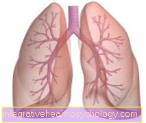
The used blood is collected in the body veins, which eventually become upper and lower Vena cava (Superior vena cava and inferior) join together and open into the right atrium. From here the blood reaches the right ventricle and is then pumped into the two lungs (see Lung). In the lungs, too, the vessels divide again down to the level of capillaries, in which the gas exchange then takes place.
The now oxygen-rich blood gets back into the heart via the two pulmonary veins (now: left atrium) and can now supply cells with oxygen again and thus gets back into the great cardiovascular system.
The sequence of the vessel sections through which the blood flows (artery-capillary-vein-heart and again from the front) is almost always followed. There are few exceptions in which a second capillary network follows before the blood returns to the heart. In this case one speaks of a portal vein system.
It happens when:
- liver
- Pituitary gland
- Adrenal gland
A jam in the pford vein system, e.g. through cirrhosis of the liver (through the scarred liver blood can no longer flow) a high pressure develops in this system, which is referred to as portal high pressure