Right atrial
synonym
Atrium dextrum
definition

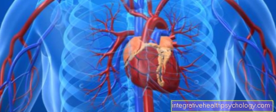
The right atrium is one of the four interior spaces of the heart that is included in the great circulation. The blood flows into it via the vena cava and is forwarded to the right ventricle.
anatomy
The right atrium is spherical and has that on the front right auricle. Of the Heart muscle is about 3 mm thick in the right atrium.
The right atrium is from the right ventricle through the right wing flap (Tricuspid valve) Cut. He gets that deoxygenated blood from the lower half of the body via the inferior vena cava; from the shoulder, chest and head regions via the superior vena cava. The common inflow path of the two veins is called Venarum cavarum sinus and represents the smooth part of the atrial wall. Within the sinus venarum cavarum there is a slight protrusion of the posterior wall of the atrium, the Tuberculum intervenosumthat delimits the venous entrances. The other part of the atrial wall is of the so-called Pectinate muscles shaped, which line the right auricle as parallel trabeculae. The boundary between these two structurally different proportions is called Crista terminalis.
The right atrium is separated from the left atrium by the atrial septum. On the right side of this partition you can see a faint indentation, Fossa ovalis. This is where there was a short circuit between the two atria during embryonic development.
The fossa ovalis is limited at the bottom by a bulge (Valvula venae cavae inferioris), which in the embryonic circulation ensures that the blood is directed through the oval fossa into the left atrium. In front of the Valvula venae cavae inferioris is the confluence of the coronary vessels, the Ostium sinus coronarii.
The right atrium also contains two important components of excitation formation and conduction: the Sinus node and the AV node.
Of the Sinus node is located next to the confluence of the superior vena cava and is considered to be primary pacemaker. This means that in healthy the Heart rate determined. He gives 60-80 pulses per minutewhich are then spread further via the heart muscle and lead to the contraction of the heart.
Of the AV node is the next station of excitation conduction. It is at the base of the interatrial septum. The AV node has one Natural frequency of 40-60 excitations per minute. Its main function is one Delay in conduction of excitationso that simultaneous contraction of the atria and ventricle is prevented. If the sinus node fails, the pacemaker function is taken over by the AV node and the heart therefore beats more slowly.
Histology - Wall Layers
Like the other internal spaces of the heart, the wall of the right atrium is made of three layers:
- Endocardium:
The endocardium forms the innermost layer and consists of one single-layer Endothelium. The function of the endocardium is one Improvement of the flow properties of the blood. - Myocardium:
The myocardium is that actual heart muscle layer and consists of two parts: the Excitation conduction system and the Working muscles. - Epicardium:
The epicardium is the outermost layer the heart wall and consists of one monolayer mesopithelium, elastic fibers and Adipose tissue. Its functions are that Compensation of the unevenness of the heart surface, Protection of the coronary arteries as well as the To facilitate volume changes of the heart.
Vascular supply and innervation
The right atrium will from the right Coronary artery provided. The venous outflow usually takes place via the Vena cardiaca parva.
The innervation of the heart is controlled by the Cardiac plexus done the fibers from the Brain stem (Vagus nerve) and from the upper thoracic Spinal cord segments contains.
function
The right atrium pumps that deoxygenated blood from the vena cava in the right ventriclewhich that Carries blood to the lungs. There the blood is enriched with oxygen (oxygenated) and brought back to the heart via the pulmonary veins to be pumped back into the body and distributed.
The contraction of the atria causes the 4. Heart sound; this can be physiological in children and adolescents, while it can be an indication of a heart disease in adults.
Clinical Aspects
The Sinus node syndrome (Sick sinus syndrome) and supraventricular extrasystoles represent diseases that originate from the atrium itself or structures of the atrium.
In which Sinus node syndrome is a Group of disordersemanating from the sinus node. These include the Sinus bradycardia, the Bradycardia-Tachycardia Syndrome, of the SA block and the Sinus node arrest.
As risk factors and causes come in high age, the coronary heart disease, Cardiomyopathies, high blood pressure or certain drugs.
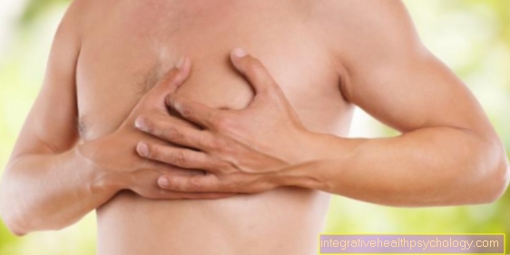
Symptoms vary depending on the type of disorder. This includes Fainting spells or dizziness, Racing heart (Pulse> 100 / min) or Chest pain.
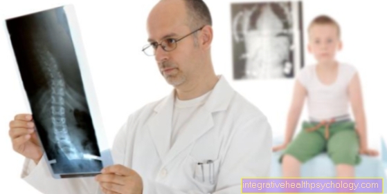
The disease can use a Long-term ECG or one Exercise ECG diagnosed and treated depending on the symptoms. At a too slow heartbeat (Bradycardia) can with the implantation of a Pacemaker be treated. At Racing heart (Tachycardia) can be appropriate Medication that lower the heart rate.
The supraventricular extrasystole, or also Palpitations called, is a disorder caused by a premature excitation of the atria is characterized. She kicks both in healthy people and in those with heart disease on, more often with age.
The triggering factors are diverse, including e.g. Inflammation, Infections, Disturbances in the salt balance, high blood pressure, the coronary heart disease, but also the Consumption of certain substances how caffeine, Drugs or alcohol.
Most supraventricular extrasystoles are mostly symptomless experienced, sometimes they are also called Heart pounding or stumbling (Palpitations) perceived.
The diagnosis is made using a EKG posed. Usually no treatment is necessary. If they occur too often or too often ß-blockers or Antiarrhythmics. In these special cases, however, it is more important to diagnose and treat the underlying disease.