Femoral neck fracture
Synonyms in a broader sense
Femoral neck fracture, Femur fracture, femur fracture, pauwels classification, Garden classification, femoral head necrosis, femoral head dying, screw connection, DHS = dynamic hip screw, hip prosthesis, osteoporosis
definition
At a Femoral neck fracture / femoral neck fracture breaks the top of the thighbone (femur) just below the head of the thigh (femur head), mostly due to a fall on the side of the hip.
causes
Older patients (geriatric patients) are predominantly affected by this form of injury. Unsteady gait and a loss of bone mass (osteoporosis) quickly lead to a femoral neck fracture after a fall. In the case of extreme bone loss, even standing up from a chair can lead to a fracture of the femoral neck. This is called a pathological spontaneous fracture.
In younger patients, considerable force is required before a femoral neck fracture occurs. Occasionally, this type of fracture looks after car accidents or fall injuries.
Appointment with a hip expert?

I would be happy to advise you!
Who am I?
My name is I am a specialist in orthopedics and the founder of .
Various television programs and print media report regularly about my work. On HR television you can see me every 6 weeks live on "Hallo Hessen".
But now enough is indicated ;-)
The hip joint is one of the joints that are exposed to the greatest stress.
The treatment of the hip (e.g. hip arthrosis, hip impingement, etc.) therefore requires a lot of experience.
I treat all hip diseases with a focus on conservative methods.
The aim of any treatment is treatment without surgery.
Which therapy achieves the best results in the long term can only be determined after looking at all of the information (Examination, X-ray, ultrasound, MRI, etc.) be assessed.
You can find me in:
- - your orthopedic surgeon
14
Directly to the online appointment arrangement
Unfortunately, it is currently only possible to make an appointment with private health insurers. I hope for your understanding!
Further information about myself can be found at
Symptoms

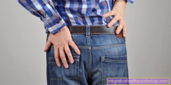
The patients usually have to be transported to the hospital by ambulance. It exists absolute inability to exercise of the broken leg. The pain at rest can be mild to unbearable. Strongest pain always pass this when trying leg to move. It is no longer possible to lift the leg from the examination table. There is tenderness and swelling on the side of the hip.
The accident event described, combined with the age of the patient and a shortened leg turned outwards, are indicative for the diagnosis by the doctor. This typical leg posture results from the displaced femoral neck fracture as well as from the corresponding muscle pull, whereby the hip external rotors predominate.

Most of the time you meet him medial femoral neck fracture and so-called pertrochanteric fractures on. Lateral femoral neck fractures are much less common.
In the case of slightly displaced femoral neck fractures, the leg position described may occasionally be slightly impressed or even absent and the patient may also experience his main complaints in the knee joint. In this case, a femoral neck fracture can be overlooked.
Injuries to larger vessels or nerves of the leg with corresponding failure symptoms are rather rare. Depending on the type of fracture, however, an interruption in the blood supply to the head of the femoral neck must be expected. However, this cannot be precisely diagnosed. Therefore, if you want to operate on the femoral head in the event of a fracture and the need for surgery, the worst situation must be assumed and the operation must be carried out as an urgent operation within 6 hours of the accident. Otherwise, the risk of femoral head death increases (Femoral head necrosis).
As an immediate measure after a femoral neck fracture, the leg is placed quietly in a foam splint, an effective pain therapy and a Thrombosis prophylaxis initiated.
As a rule, we nowadays do without an extension (pull on the leg to counteract the shortening).
diagnosis
This is decisive for the final confirmation of the suspected diagnosis of a femoral neck fracture / femoral neck fracture X-ray image. A pelvic overview image and an axial image of the hip are usually taken. In the vast majority of cases, no further diagnostic imaging is necessary afterwards.
In young patients who have been exposed to considerable violence, the diagnosis is usually carried out by a Computed Tomography (CT) or Magnetic resonance imaging of the hip (MRI of the hip) to cover other injuries (e.g. hip socket fractures, Pelvic fractures etc.) to be recorded and specified.
classification

Classifications serve the communication between specialists and allow a derivation of medical therapy measures, which are summarized in recommended guidelines for the individual specialist areas.
Common classifications of the different types of fractures for a femoral neck fracture are those according to Pauwels and Garden.
The Pauwels classification is based on the inclination of the fracture surface.
- Pauwels I: fracture surface <30 ° to the horizontal plane
- Pauwels II: fracture surface 30 ° -70 ° to the horizontal plane
- Pauwels III: fracture surface> 70 ° to the horizontal plane
The smaller the fracture surface angle, the more stable the fracture. As the fracture surface angle increases, the risk of a false joint increases.
The Garden classification is based on the position of the femoral head.
- Garden I: Valgisch (stable) indented fracture
- Garden II: Unmoved break
- Garden III: Varisch (unstable) indented fracture
- Garden IV: Strong fracture displacement
The risk of femoral head death increases with the number of Gardeners.
Therapy of femoral neck fracture
A femoral neck fracture / Femoral neck fracture must be surgically treated in the vast majority of cases. The fracture is seldom so stably indented that conservative treatment is possible. But even if a femoral neck fracture is stable, for most older patients a 3-month relaxation period for the leg is out of the question. The resulting immobilization leads in many cases to life-threatening complications such as one
- lung infection
- Leg vein thrombosis or
- Pulmonary embolism.
Therefore, conservative therapy is reserved for the rare young patients who can also be mobilized with complete relief of one leg.
In principle, a distinction is made between the femoral head received or replacing Operations. Operations to preserve the femoral head should be carried out as quickly as possible (within 6 hours of the accident) in order to prevent the risk of death of the femoral head.
Treatment options that preserve femoral head are:
- Screw connection: Three screws are screwed through the Femoral neck of Thighbone introduced into the femoral head. The cartilaginous surface of the femoral head is not broken through. The screws should be as parallel to each other as possible and the screw thread should not cross the fracture line so that the femoral neck fracture can collapse under load.
Advantage: quick operation. Little Soft tissue injury. Femoral head and thus the natural one hip joint remain.
Disadvantage: If the bone substance is poor (osteoporosis) Slipping of the fracture or false joint formation (Pseudarthrosis) possible. Immediate full load is not possible.
- Dynamic Hip Screw (DHS): One Metal plate screw construction is attached to the thigh. The screw runs through the femoral neck into the femoral head and has the ability to slide like a telescope, creating a compression effect in the fracture area.
Advantage: quick operation. The femoral head and thus the natural hip joint are retained.
Disadvantage: the fracture may slip off. Immediate full load is not possible. Frequent femoral head necrosis.
- artificial hip joint: In geriatric patients with worse Bone substance, pre-existing hip osteoarthritis and foreseeable difficulties in mobilization can primarily be the implantation of a Hip prosthesis be displayed.
Advantage: Immediate pain-adapted full weight bearing possible. Easier early mobilization. No slipping of the fracture or death of the femoral head possible.
Disadvantage: major surgery. Greater soft tissue trauma. Replacement surgery necessary if the prosthesis loosens.
Complications
Complications of surgical treatment of the femoral neck fracture:
- Vascular, tendon and nerve injuries
- Thrombosis / pulmonary embolism
- infection
- Slipping of the fracture
- Implant loosening
- False joint formation (pseudarthrosis)
- Femoral head necrosis
Follow-up treatment / prognosis
Postoperative early mobilization is essential for the mostly older patients. That is why mobilization begins on the first postoperative day by standing on the bed. In the following period, the operated leg may only be partially loaded (15-20 kg) for a DHS for a period of 6-12 weeks. The introduced metal (osteosynthesis material) can be removed after one to two years, possibly not at all. The full load is reached after approx. 3 months. Regular x-ray controls document the progressive healing of the fracture.
When a hip prosthesis is implanted, the load can be increased immediately. Depending on the choice of implant and bone substance, pain-dependent full loading is sometimes possible immediately.
A fracture of the femoral neck can also have some long-term consequences.
Read more on this topic at: Femoral neck fracture late effects and Pain after hip surgery
Remove screws
After an operative treatment of the femoral neck fracture, the implant removal, i.e. the removal of the Osteosynthesis material (Screws), not absolutely necessary in all cases.
Certain screw systems can also remain in the bone.
A high patient age can also be a reason for leaving a screw. Overall, in addition to the age and choice of implant, the patient's level of activity and possible local discomfort in the hip region influence the decision to remove the screws.
In most cases, however, an implanted screw system will work after about 2 years away.
The screws are removed if during an inpatient stay. This is an operation that requires greater exposure and the risk of complications is too high to be performed on an outpatient basis.
Removal of the implant is necessary because if left in place there may be an increased risk of certain complications. It can for example too Fatigue fractures of the implant or infections.
In addition, endoprosthetic treatment of any other injury in the vicinity of the implant can be difficult.
Ultimately, there may be adhesions in the implant, so that the time for screw removal must be chosen in good time.
As with almost every surgical procedure, the risks involved in removal include the risk of nerve, vascular and soft tissue damage. Heavy bleeding and infections can also occur. An X-ray check must be carried out after each removal to ensure that no screw residues have remained and that no new fractures have occurred as a result of the explantation.
It is very important to appear for regular wound control and to try to restore full resilience and functionality in the form of physiotherapy. As with the treatment of the femoral neck fracture with osteosynthesis such as the screw after the operation, the patient should be mobilized quickly.
To support this, patients are prescribed physiotherapy. Since after such an operation the risk of thrombosis is increased, each patient receives a drug for a certain period of time Thrombosis prophylaxis.
Duration
Compared to the length of stay after surgery for first aid (several weeks) patients can leave the hospital after a few days if the screws are removed without complications.
The aim is to achieve full weight-bearing as soon as possible, but patients should use forearm crutches to relieve the pressure in the first few days.