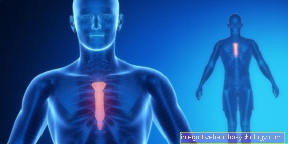
The chest breathing
definition
The chest breathing (Chest breathing) is a form of external breathing. It is used to exchange breathable air by ventilating the lungs (ventilation). With chest breathing, this ventilation takes place by expanding and contracting the chest.
With this form of breathing, the ribs rise and fall visibly, and they also give way to the outside. Your movements come from the tension (contraction) and relaxation of the intercostal muscles.
A mixture of chest breathing and the other form of breathing, abdominal breathing, is usually used unconsciously.
Find out more at: Human breathing

How does chest breathing work?
The chest breathing (Chest breathing) is used for external breathing and thus for the exchange of breathable air. In contrast, internal respiration is a form of energy production at the cellular level.
More information can be found here: Cellular respiration in humans
External breathing is used to supply the body with vital oxygen. At the same time, carbon dioxide, which is produced when the cells generate energy, is released into the environment. The exchange of breath takes place in the lungs. The lungs must always be adequately ventilated so that it can run off without problems.
With chest breathing, this happens through the interplay of expanding and contracting the chest. ly the ribs and the intercostal muscles are involved (Intercostal muscles). When there is an increased need for oxygen or acute shortness of breath, the so-called auxiliary respiratory muscles also support the thorax movement.
- On inhalation (inspiration) the external intercostal muscles (External intercostal muscle) together. This lifts the ribs and turns them outwards. The thorax expands. Since the lungs are over the pleura (Pleura) is connected to the rib cage, it follows this movement. The lungs also expand, their volume increases. This creates a negative pressure. More air now flows through the airways into the lungs to compensate for the negative pressure. This is where the actual inhalation takes place.
- The exhalation (Expiration) is possible with normal, not strained breathing without supporting muscles. The lungs have a so-called inherent elasticity. This means that it consists of tissue that strives to contract as much as possible. If the outer intercostal muscles relax, the lungs are no longer held wide. It follows its own elasticity and contracts. This creates an overpressure that drives the air out of the lungs. So there is exhalation.
Normal, unconscious breathing consists of a mixed form of chest breathing and abdominal breathing.
Chest breathing disorders
Chest breathing can be a result of illness unnaturally strong or frequent occur.
- Is it difficult to breathe (Dyspnea), the proportion of chest breathing increases and that of abdominal breathing decreases. In case of severe shortness of breath (Orthopnea) the auxiliary respiratory muscles are also used. People who suffer from orthopnea, often sit upright, have the Arms propped up and breathe hard. Such shortness of breath can be triggered by a wide variety of factors. On the one hand due to diseases of the lungs such as bronchial asthma, the chronic obstructive pulmonary disease COPD, pulmonary embolism or pneumonia (Pneumonia). But also heart problems such as heart failure (Heart failure), Heart valve defects or heart attacks can lead to it.
Learn more about: Diseases of the lungs
- Is the Abdominal breathing impaired, increased chest breathing takes over its function. This can e.g. in the case of swelling of the liver or spleen, but also in pregnancy or very overweight (Obesity) be the case.
- The increased chest breathing can also be a sign of psychological problems. It occurs, for example, during fast, deep breathing (hyperventilation). This can be a sign of Panic attacks or Anxiety disorders be. Chest breathing also increases in some cases with depression. Since chest breathing is mainly used by the body when there are high demands, such as stress, it can also be a sign of a high level of stress. There too strong pain Cause stress, chest breathing increases in this case as well.
- Chest breathing can also be directly affected by an illness. This is the case, for example, when the muscles in the chest that are required for chest breathing are very tense. Excessive strain on chest breathing when there is a lot of stress can overstrain the muscles and make them tense. In addition, you can Skeletal malformations, Bad posture and Sedentary lifestyle lead to tension. These can sometimes be very painful and even to one Feeling short of breath to lead. Targeted exercise, strengthening the muscles and relaxation techniques help in these cases.
- This form of breathing is also restricted if the muscles involved in chest breathing are damaged. This is how muscle weaknesses spread (Muscular atrophies) also work on these muscles.
- A muscle can also recede if the nerve that normally supplies it fails. The main muscles of chest breathing, the external intercostal muscles, are supplied by many nerves (nervi intercostales). If only one fails, the neighboring nerves take over supplying the affected muscles. However, if several nerves are affected, breathing problems can occur.
You might also be interested in: Pain on inhalation
What is diaphragmatic breathing?
As diaphragmatic breathing (Diaphargical breathing, Abdominal breathing) is called a form of breath. It is characterized by the contraction and relaxation of the diaphragm. During diaphragmatic breathing, the abdominal wall rises and falls visibly.
The diaphragm contracts to inhale. This shifts it downwards. The pleura that has grown together with it follows this movement. This creates a negative pressure in the space between the lungs and the diaphragm. Following this negative pressure, the lungs expand and air flows in.
The lungs constantly strive to contract (inherent elasticity). Following this inherent elasticity, it decreases again as soon as the diaphragm relaxes. The diaphragm moves upwards in the body. Abdominal breathing did most of the breathing at rest. It is supported by the second form of breathing, abdominal breathing.
You can find detailed information on our main page: The diaphragmatic breathing
What is the difference to abdominal breathing?
There are two types of breathing, chest breathing and abdominal breathing. Both forms take place in normal breathing at rest. Abdominal breathing predominates.
On the one hand, the two types of breathing differ in the muscles involved. Chest breathing is mainly done by the intercostal muscles, with higher demands being connected to auxiliary breathing muscles in the shoulder, neck, back and abdominal area. The negative pressure in the space between the pleura and the lungs, which is necessary for inhalation, is achieved here by expanding the chest.
During abdominal breathing (Diaphragmatic breathing) is mainly the diaphragm (Diaphragm) active. Breathing can be supported here by the abdominal muscles. During abdominal breathing, the negative pressure arises from the fact that the diaphragm contracts and shifts downwards in the body.
The two forms of breathing also differ in their energy consumption:
- Abdominal breathing requires less energy because fewer muscles are active.
- In addition, chest breathing is more likely to be used during tension and activity.
- Abdominal breathing dominates in rest and relaxation. This is why it can be relaxing to breathe deeply into your stomach. Abdominal breathing is also easier to control than chest breathing. That is why she plays a role, for example Singers and Musicians, but also with some Martial arts, a major role.
Which muscles are involved in chest breathing?
The muscles involved in chest breathing are muscles of the so-called skeletal muscles. The muscles can thus be controlled arbitrarily. The muscles involved in breathing are divided into respiratory and auxiliary respiratory muscles. The separation of these two muscle groups is not always sharp.
The auxiliary respiratory muscles are used to increase breathing Shortness of breath or increased oxygen demand.
Chest Breathing Muscles
The following muscles belong to the respiratory muscles:
- The external intercostal muscles (External intercostal muscles) are involved in breast augmentation even at rest. They run diagonally between the ribs and can lift them and turn them outwards. They are used for inhalation (inspiration). The exhalation normally takes place without muscle support.
- During exertion with increased energy requirements and higher levels of carbon dioxide, however, exhalation should be accelerated. The inner intercostal muscles serve this purpose (Musculi intercostales interni, Musculi intercostales intimi). These muscles also run obliquely between the ribs, but in the opposite direction to the outer intercostal muscles.
- The subcostales muscle, which is derived from the internal intercostal muscles, also fulfills this function.
- In addition, the transversus thoracis muscle, which runs from the back of the sternum to the ribs, supports exhalation.
Muscles of the auxiliary respiratory muscles
The auxiliary respiratory muscles are divided according to whether they support inhalation or exhalation.
- At the breathe in The pectoralis major muscle helps in particular.
- The pectoralis minor muscle also has this function.
- The serratus anterior muscle, which runs from the ribs to the shoulder blade, also assists inhalation. These muscles usually pull the shoulder girdle towards the ribs and up the rib cage. If the shoulder girdle is fixed, e.g. by propped up arms (e.g. in the driver's seat), the muscles can use all their strength to lift the chest. As a result, they support inhalation in the driver's seat even more.
- Neck muscles can also help with inhalation. For example the sternocleidomastoid muscle, which runs from the sternum to the collarbone and to the head. When contracting, it can lift the chest.
- The scalene muscles, which can be divided into posterior, anterior and middle muscles, have the same function. They move from the cervical vertebrae to the ribs.
- In addition, the serratus posterior superior back muscle is used for assisted inhalation. It pulls from the cervical and thoracic vertebrae to the ribs.
- In a broader sense, the muscle cord that runs along the spine and helps to straighten up (erector spinae muscle) is also counted among the auxiliary respiratory muscles that support inhalation.
Muscles used for exhalation
Exhalation is mainly supported by the abdominal muscles. This is also known as the so-called. Abdominal press. They include:
- The rectus abdominis muscle and the transversus abdominis muscle, which also form the abdominal wall, are involved in exhalation. In addition, the musculus obliquus internus abdominis and the musculuc obliquus externus abdominis belong to these muscles. They run diagonally along the side of the stomach.
- The quadratus lumborum muscle, which can fix the 12th and thus the last rib, also supports expiration.
- In addition, two back muscles help with exhalation: one is the serratus posterior inferior muscle, which pulls from the spine to the lower ribs.The latissimus dorsi muscle, which runs in the shape of a triangle over the lower back to the shoulder blade, also has this function. It is particularly active when coughing and is therefore also known as the cough muscle.
Recommendations from our editorial team
- Breathing exercises
- Human breathing
- Stinging of the heart when inhaling
- Lung pain
- Diseases of the lungs


