meiosis
definition
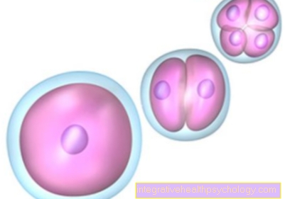
Meiosis is a special form of nucleus division and is also known as meiosis. It contains two divisions that turn a diploid parent cell into four haploid daughter cells.
These daughter cells each contain a 1 chromatid chromosome and are not identical. These germ cells are needed for sexual reproduction.

introduction
In men, the germ cells are the sperm cells formed in the testes. The equivalent in women are the egg cells she has from birth.
One haploid germ cell from each parent merges into a double set of chromosomes, which is found in all other body cells.
If one of the two divisions is faulty during meiosis, numerical chromosomal aberrations, such as trisomy 21 (known as Down syndrome).
What is the function of meiosis?
The function of meiosis is the production of germ cells in both female and male living things. These are required for sexual reproduction and are accordingly found in mammals and humans.
After meiosis, a cell with a double (diploid) Chromosome set of four cells with a simple (haploid) Set of chromosomes.
This reduction in the set of chromosomes is very important, as otherwise two germ cells with a double set of chromosomes would fuse together during fertilization. The result would be a living being with four times (tetraploid) Set of chromosomes. This chromosomal abnormality accounts for approximately 5% of all miscarriages.
In addition to reducing the number of chromosomes and producing germ cells, meiosis has another function. By randomly distributing the chromatids to the four daughter cells, meiosis ensures genetic diversity.
In addition to the random distribution of the genome, there is also the exchange of genetic information between maternal and paternal chromosomes. This process is known as crossing-over and increases genetic recombination and diversity again.
This topic may also be of interest to you: Tasks of the cell nucleus
What is the course of meiosis?
The course of meiosis is always the same and can be roughly divided into two parts. These in turn consist of several phases, which, however, are identical in both divisions.
First division of meiosis
Meiosis starts with the doubling of the two chromatids, so that the cell has a double set of chromosomes with four chromatids. This is followed by the first division of meiosis, in which the two pairs of chromosomes are separated from one another.
The two resulting cells each have a chromosome with two chromatids. This division is known as a reduction division, as the double set of chromosomes is halved. It runs in several phases, which have the same names as in mitosis:
- Prophase
- Metaphase
- Anaphase
- Telophase.
In addition, in this part of meiosis, the genetic material is still recombined within the chromosomes. It is an exchange of certain sections of DNA between the two chromosomes, which is known as crossing-over.
Second division of meiosis
The second part of meiosis consists of the so-called equation division. Here the two sister chromatids are separated from each other. A total of four germ cells are created which, as genetic material, only contain one chromatid.
As in the first meiotic division, the four phases (Prophase, metaphase, anaphase, telophase) find again.
The separation of the sister chromatids in the second part of meiosis can be compared to mitosis, since there the chromatids are separated and drawn to opposite cell poles.
What are the phases of meiosis?
Meiosis is important for germ cell development and can be divided into different stages. First of all, one must differentiate between meiosis I and meiosis II. This division makes sense because two cell divisions occur during meiosis.
The first division is called the reduction division because the two homologous chromosomes are separated from each other. This creates a single set of chromosomes from a double set of chromosomes.
This first meiosis can be divided into four phases:
- Prophase I
- Metaphase I.
- Anaphase I.
- Telophase I.
The original cell has two chromosomes that are duplicated by replication. The result is a cell with four chromatids.
In prophase, the chromosomes are compressed and approach each other. This spatial proximity of both chromosomes is important for the following crossing over. Both chromosomes exchange genetic material, which increases genetic diversity.
Next comes the metaphase, in which the two homologous chromosomes are arranged in the equatorial plane. At the same time, the spindle apparatus is formed.
In the anaphase, the chromosome pairs are separated from each other and drawn to the opposite cell poles.
In the last phase, the telophase, the cell membrane constricts so that two daughter cells are created. These have a simple set of chromosomes, but it consists of two chromatids.
Next comes the second division of meiosis. This is called equation division and affects both haploid daughter cells. During this division, the sister chromatids are separated from one another, so that a total of four cells with one chromatid result.
Meiosis II is very similar to mitosis and can also be divided into the same phases:
- Prophase II
- Metaphase II
- Anaphase II
- Telophase II
In the prophase the sister chromatids condense and the spindle apparatus begins to form.
In the metaphase, the chromatids are arranged in the equatorial plane so that both chromatids are approximately the same distance from the cell pole.
In the anaphase, the sister chromatids are separated from each other and migrate towards the cell pole.
In the telophase the cell membrane constricts again and new nuclear envelopes are formed.
This resulted in a total of four daughter cells that contain a simple set of chromosomes in the form of a chromatid as genetic material.
These germ cells, sex cells or gametes, are created differently in both sexes.
In women, the egg cells are present from birth, but are in a kind of dormant mode until puberty. With the onset of sexual maturity, an egg cell matures every month, which can then be fertilized.
In men, the production of sperm in the testicles does not begin until the onset of puberty. In contrast to women, men are still able to form germ cells well into old age.
What is the difference to mitosis?
Meiosis is very similar to mitosis with regard to the second meiotic division, but there are some differences between the two cell nucleus divisions.
- Result
The result of meiosis are germ cells with a simple set of chromosomes that are suitable for sexual reproduction. In mitosis, identical daughter cells with a double set of chromosomes are created. These cells do not have the function of reproduction, but replace old, dead or no longer fully functional body cells.
- Number of divisions
Another difference between meiosis and mitosis is the different number of divisions. Two divisions are necessary in meiosis. In the first reduction division, the two pairs of chromosomes are separated; in the following equation division, the two sister chromatids are separated from each other.
In contrast, with mitosis, one division is sufficient. In this one division, the sister chromatids are separated so that two genetically identical daughter cells arise.
- Duration
However, meiosis and mitosis differ not only in their function and the number of divisions, but also in their duration. Mitosis is a relatively quick process that takes about an hour to complete. Meiosis, on the other hand, takes a lot longer and can stagnate in one phase for several years or even decades.
This is the case with the egg cells that are already created at birth, but are all in a sleep mode until they reach sexual maturity.
The development of male sex cells, the sperm, also takes around 64 days. Of these, about 24 days fall to meiosis.
You can find more information on this topic at: Mitosis - Simply Explained!
What is the crossing over?
The crossing-over describes the exchange of genetic material between two chromatids. In the process, these approach each other, cross over and then exchange certain DNA fragments.
This process takes place during germ cell division (meiosis) instead of. The crossing-over can be assigned to prophase I, which can be divided into five phases.
Before the first division of meiosis begins, the DNA is doubled so that there are four chromatids in the cell. The first phase of prophase I is that Leptotene, in which the chromosomes condense and become visible under the light microscope.
This is next Zygotene, in which the chromosomes approach each other and a homologous chromosome pairing is created. This spatial proximity of both chromosomes is the prerequisite for being able to exchange genetic material. In parallel, the synaptonemal complex is formed. This is a complex of several proteins that is formed between the chromosomes and ensures the exact position of these.
Hereinafter Pachytan the actual crossing-over now takes place. There were already breaks in the DNA in the two previous phases. Now two chromatids cross each other and the break points are repaired. The maternal and paternal chromosomes exchange small segments of DNA. These crossovers are visible as chiasmata under the light microscope.
in the Diploma the synaptonemal complex dissolves and the chromosomes are only connected to one another at the chiasmata.
In the last phase of prophase I, the Diakinesis, the nuclear membrane dissolves, the mitotic spindle begins to build up and meiosis can take place in the usual sequence.
The crossing-over is used for intrachromosomal recombination and, together with the random assignment of the genetic material to the germ cells, plays an important role in the variety of characteristics.
How does trisomy 21 occur?
Trisomy 21 is a disease caused by the triple presence of the 21st chromosome. In healthy cells there are duplicate chromosomes, so that humans have a total of 46 chromosomes. A patient with trisomy 21 has 47 chromosomes and has Down syndrome.
The threefold presence of the 21st chromosome is caused in most cases by an error in meiosis.
Meiosis turns a cell with 4 chromatids into four germ cells with one chromatid each through two divisions. However, errors can occur in both maturities. As a result, the chromatids are not correctly distributed over the germ cells and germ cells with two chromatids follow.
These germ cells have duplicate individual chromosomes, in Down syndrome the 21st chromosome. If such an egg cell fuses with a healthy sperm, the embryo has the 21st chromosome in three copies.
The symptoms of this numerical chromosomal aberration are diverse and can vary in severity. Often such children are spiritually retarded, stunted and have congenital heart defects.
The risk of disease increases with the age of the mother, so that many older pregnant women prefer to have an amniotic fluid test carried out.
If you have any further interest in this topic, then read our next article below: Down syndrom
Recommendations from the editorial team
Further general information may also be of interest to you:
- Mitosis - Simply Explained!
- Chromosome set - how is it in humans?
- Chromosome mutation - which ones are there?


.jpg)