Intestines
Structure of the intestine
Life is not possible without the gut. The vital digestion is controlled and guaranteed through it. The food and the fluids enter the human body via the intestine, and this is where the breakdown into usable and non-usable ingredients of the food takes place. The human intestine is divided into numerous sections, each of which has different tasks and parts in the digestive process.
The main division is the distinction between Small intestine and Large intestine. To the stomach the small intestine joins with all of its sections. One distinguishes the Duodenum (Duodenum), which connects directly to the stomach outlet. In him the Bile acids of the Gallbladderwhere they are stored, to the food that is already sufficiently reduced in size and mixed with fluid in the duodenum. Rather, it is now a chyme that pushes its way through the tight intestinal plexus with rhythmic muscle movements. Chemical digestion of the chyme begins with the mixing of bile acids. Of the pancreas produced, enzymes reach the small intestine, which break down the various fats. That would be mentioned here Lipase and the Amylase as the most important enzymes. This is attached to the duodenum Jejenum on. It makes up about 40% of the small intestine.
The remaining 60% are from the so-called Ileum educated. The main task of these sections of the small intestine is to knead the chyme and absorb nutrients. So be next to the need Nutrients also Folic acid, vitamin C and Calcium withdrawn from the chyme in the small intestine.
Since food is contaminated with bacteria to a not inconsiderable extent, a large part of the is located human immune and defense system in the intestine, in order to quickly eliminate the corresponding pathogens and intruders. The immune system is created in the form of lymphatic structures. The optimal absorption of the nutrients is achieved by a mucous membrane that spreads in waves and slips the entire inner wall of the small intestine. The Villi protrude into the intestinal lumen and thus come into contact with the chyme that is pushed through the intestine. Shortly after the duodenum, the villi are largest; the further the intestine descends, the flatter they become. They are almost invisible up to the colon. The small intestine takes up a large area, which is also enlarged by the ingenious folds. It also offers a large target for diseases. Common bowel diseases can autoimmune be and will be as Ulcerative colitis or Disease Crohn designated. Symptoms are severe diarrhea, at times with blood and Convulsions socialized.
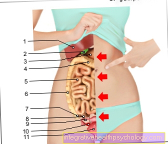
Illustration of the digestive tract

Digestive tract
A. - Food route
a - digestive organs
in the head and neck
(upper part of the digestive tract)
b - digestive organs
in the body cavity
(lower part of the digestive tract)
- Oral cavity - Cavitas oris
- Tongue - Lingua
- Sublingual salivary gland -
Sublingual gland - Trachea - Trachea
- Parotid gland -
Parotid gland - Throat - Pharynx
- Mandibular salivary gland -
Submandibular gland - Esophagus - Esophagus
- Liver - Hepar
- Gallbladder - Vesica biliaris
- Pancreas - Pancreas
- Colon, ascending part -
Ascending colon - Appendix - Caecum
- Appendix -
Appendix vermiformis - Stomach - Guest
- Large intestine, transverse part -
Transverse colon - Small intestine - Intestine tenue
- Large intestine, descending part -
Descending colon - Rectum - Rectum
- Nach - anus
You can find an overview of all Dr-Gumpert images at: medical illustrations

To the Small intestine closes the Large intestine which is also called colon referred to as. Here there is no longer the anatomical folds of the mucous membrane that protrude into the chyme. The walls are flatter and smoother, and a large part of the nutrient utilization has already been completed in this section of the digestive tract. The large intestine begins at an anatomical structure that strictly separates the small intestine from the large intestine. This structure is also called the Bauhinsche flap designated. It follows the appendix (appendix), which is found in the lower right part of the abdomen in most people. If it was previously believed that this section of the intestine had no essential tasks, today it is known that a large part of the immune response occurs in the appendix. Most people are familiar with this section of the intestine, possibly from their own experience, because the Spinous process the appendix can become inflamed and must then be surgically removed in most cases.
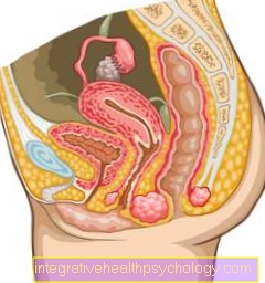
You can find more information about the anatomy of the abdominal cavity here: Abdomen
Figure large intestine

- Colon, ascending part -
Ascending colon - Appendix - Caecum
- Appendix -
Appendix vermiformis - Right colon bend -
Flexura coli dextra - Large intestine, transverse part -
Transverse colon - Left colon bend -
Flexura coli sinistra - Large intestine, descending part -
Descending colon - Large intestine, s-shaped part -
Sigmoid colon - Rectum - Rectum
- Bulges of the
Colon Wall -
Haustra coli - Liver - Hepar
- Stomach - Guest
- Spleen - Sink
- Gallbladder -
Vesica biliaris - Small intestine -
Intestine tenue - Esophagus -
Esophagus
You can find an overview of all Dr-Gumpert images at: medical illustrations
Illustration small intestine

- Small intestine -
Intestine tenue - Duodenum, upper part -
Duodenum, pars superior - Duodenal
Jejunum junction -
Duodenojejunal flexure - Jejunum (1.5 m) -
Jejunum - Ileum (2.0 m) -
Ileum - End part of the ileum -
Ileum, pars terminalis - Colon -
Intestinum crassum - Rectum - Rectum
- Stomach - Guest
- Liver - Hepar
- Gallbladder -
Vesica biliaris - Spleen - Sink
- Esophagus -
Esophagus
You can find an overview of all Dr-Gumpert images at: medical illustrations
By definition, the actual large intestine begins (colon) just behind the appendix. One distinguishes one with the colon ascending part (Pars ascendens), one transverse part (Pars transversum) and one descending part (Pars descendens). Viewed from the front, the large intestine forms a kind of frame in the middle of which the small intestine is embedded. Viewed from the outside, the large intestine is characterized by constrictions, also known as House doors are designated. Its main task is the absorption of minerals and the removal of water from the chyme. In total, the large intestine can process 300 ml of chyme into 150 g of stool. In addition to the absorption of important minerals, substances are also released into the intestine and thus made to be excreted. Above all, that should be mentioned here potassium and the Bicarbonate, which has important buffering functions and is excreted via the intestines in the event of increasing alkalinization of the body. Numerous toxins are also ultimately eliminated via the large intestine and thus leave the body unnoticed. The intestine is never sterile and is flooded with numerous bacteria that are part of the intestinal flora. The task of the numerous bacteria are defense against pathogens through a natural barrier function, support of the metabolism in the colon mucosa and acceleration of exchange processes (exchange of nutrients etc. on the intestinal wall). They also stimulate bowel activity and the mechanical movement of the bowel. In addition, the stimulation of the immune system is counted among the tasks of the bacteria. Through the Intestinal flora an optimal environment is maintained in the intestine, which maintains the exchange processes of nutrients and pollutants. A disproportion of the intestinal flora leads to the overturning of the balance and ultimately to Diarrhea. Often this is after a long time Taking antibiotics to observe.


.jpg)