Choroid plexus
What is the choroid plexus?
The choroid plexus is a collection of intertwined blood vessels. Both veins (running towards the heart) and arteries (running away from the heart) are involved in the formation of the plexus.
They are all located in cavities inside the brain (cerebral ventricles) that are filled with cerebrospinal fluid (liquor). The function of the choroid plexus is to form the liquor and deliver it to the ventricles.
Read more on the topic: Cerebral ventricle

Anatomy of the choroid plexus
The choroid plexus is composed of two layers. The inner layer (lamina propria) consists of a specialized form of the soft meninges (pia mater). Richly branched, tiny blood vessels (capillaries) can be found in it. The capillaries represent the transition between veins and arteries. The outer layer (lamina epithelialis) also consists of specialized supporting cells of the nerve tissue. This special type of cell is known as ependymal cells. They filter the blood from the inner layer and thus produce the cerebrospinal fluid (liquor).
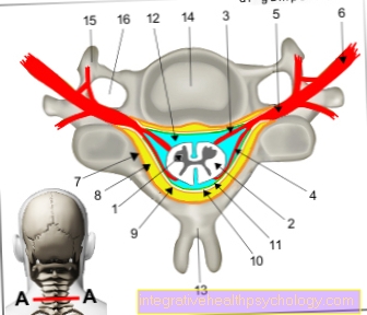
There are several choroid plexuses. They lie in liquor-filled cavities inside the brain (cerebral ventricles). There are 4 cerebral ventricles. The first two (lateral ventricles) lie next to each other, one in each half of the brain. The third and fourth ventricles lie one below the other under the lateral ventricles. The spinal canal (central canal) connects to the fourth ventricle. The cavities are connected to one another by holes and small passages.
The choroid plexus is found in the lateral ventricles, especially on the inside on the underside. In the third and fourth ventricles it is more likely to be located on the upper side. The fourth ventricle has a special feature: on its sides there are small holes (lateral aperture, Luschkae foramen). Part of the choroid plexus exits through these holes. Because of its shape, this structure is known as Bochdaleck's flower basket.
Find out more about the topic: Liquor (cerebrospinal fluid)
Function of the choroid plexus
The task of the choroid plexus is to form the Gehrin spinal cord fluid (liquor cerebrospinalis). It produces around 500 ml of liquor per day. In this way, the choroid plexus renews the entire cerebral fluid several times a day. The cerebrospinal fluid is vital for the brain. The brain lies in it as if it were floating in water. This will protect it from bumps. In addition, the buoyancy of the liquor reduces the weight of the brain. This also prevents injuries caused by the effects of pressure.
Another important function of the CSF is the disposal of waste products from the nerve cells of the brain. During the metabolism of the nerve cells, substances accumulate that they can no longer use. They are released into the liquor. This transports them with its direction of flow into the lymphatic system. The choroid plexus ensures that there is enough cerebrospinal fluid to perform these tasks.
It produces the liquor by filtering the blood from the capillaries of its inner layer. The fluid in the blood (the blood plasma) is separated from the solid components of the blood (the blood cells). The ependymal cells of the choroid plexus release other substances into the fluid obtained in this way, e.g. Sodium, magnesium, chloride, glucose and vitamins. This leads to an increased concentration of these substances in the liquor and serves to optimally supply the nerve cells.
Disorders of the choroid plexus
Cysts
A choroid plexus cyst is a cyst in the tissue of the choroid plexus. Cysts are closed, newly formed cavities in an organ. They are found in the choroid plexus almost only in unborn children. They can occur individually or be in several places. As a rule, they are only a few millimeters in size.
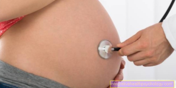
In the vast majority of cases, the cysts do not cause any problems. They occur relatively frequently in 1-2: 100 children. In the course of pregnancy (up to the 28th week of pregnancy) they usually resolve on their own. Most often, the choroid plexus cysts are noticed during an ultrasound examination (sonography) of the pregnant woman and the child. Such a finding can lead to great uncertainty and concern.
Choroid plexus cysts are not a disease, however. The cyst by itself does not affect the child's brain development prior to birth. In extremely rare cases, the cysts can be so inconvenient that they block the drainage of the CSF and the CSF collects in the child's head (hydrocephalus internus). This rare complication usually only occurs after birth and can be treated.
Find out more about the topic: Brain cysts
If the choroid plexus cysts occur during an otherwise normal pregnancy, they are usually completely harmless. From a statistical point of view, however, they are associated with an increased risk of a chromosome number disorder (chromosome aberration). Especially the risk of trisomy 18 (Edwards Syndrome), i.e. the presence of three chromosomes 18, is increased in this case. This risk increases slightly if the mother is over 35 years old or if the choroid plexus cysts appear on both sides. Therefore, a more detailed ultrasound examination (fine ultrasound) of the child should be carried out.
In addition, the presence of the cysts should be checked over the 28th week of pregnancy. Invasive diagnostics (amniocentesis / amniocentesis or chorionic villi biopsy) can also be used to rule out chromosome aberration. With these examination methods, the amniotic fluid or part of the placenta is punctured. This increases the risk of miscarriage by up to 2%. If the results are normal, the risk of having a child with a chromosome abnormality is significantly lower. Therefore, such an examination should be considered very critically if the findings are normal.
If the findings are conspicuous, it is possible to seek advice on invasive diagnostics. This should be done by a human geneticist or a doctor with appropriate training. The individual risk should be calculated and explained during the conversation.
For more information, see: Amniotic fluid examination
tumor
Tumors in the choroid plexus can be both benign (benign) and malignant (malignant). The benign form is called plexus papilloma, the malignant form plexus carcinoma. In 80% of cases, a choroid plexus tumor is a papilloma plexus. Tumors of the choroid plexus usually occur in infancy or childhood, later they become significantly less common. The tumor often produces cerebrospinal fluid. It can also clog the CSF drainage path. This leads to a build-up of fluid in the brain called hydrocephalus. This can lead to increased pressure on the brain and other symptoms such as headache, nausea, vomiting and seizures.
For more information, see: Head of water in the baby
The diagnosis is made using imaging techniques such as computed tomography, magnetic resonance imaging, or a biopsy of the tumor. The therapy consists in a possible complete microsurgical removal of the tumor, possibly followed by radiation therapy. With plexus papilloma, the chances of survival after therapy are good. The tumor rarely metastasizes or cannot be completely removed. Plexus carcinoma, on the other hand, often metastasizes. The prognosis is therefore unfortunately not favorable.
calcification
Calcification of the choroid plexus is the term used to describe the deposition of solid matter in the area of the choroid plexus. It doesn't have to be lime, proteins can also lead to this picture. Calcifications are usually noticed as an incidental finding in imaging procedures such as magnetic resonance tomography or computed tomography.
Since calcification occurs in a relatively large number of people, especially in older age, it is currently assumed that it has no disease value. In some cases, the calcification can indicate vascular calcification of the vessels of the brain (arteriosclerosis) or minor trauma. An increased incidence of brain tumors has also been observed very rarely.
Recommendations from the editors for you
- Blood-brain barrier
- Cerebral ventricle
- Meninges
- Cerebrum