Foot muscles
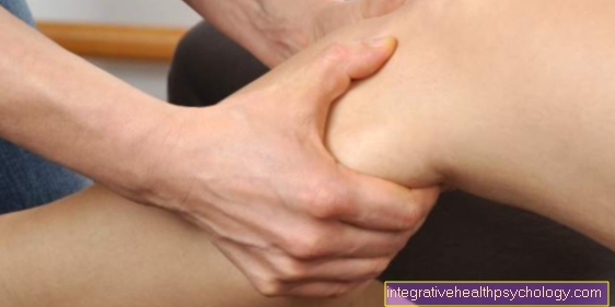
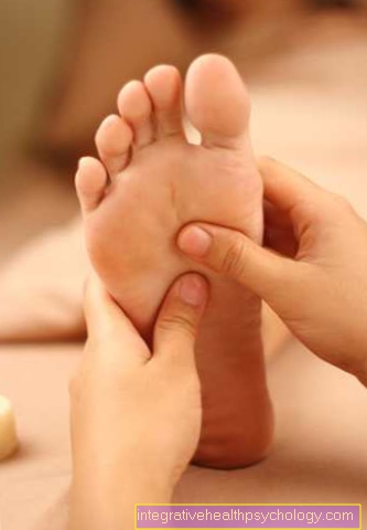
The foot muscles are divided into the muscles of the back of the foot (dorsum pedis) and the sole of the foot (planta pedis). They enable the foot to move in various dimensions and are also involved in its stabilization.
The foot muscles are divided into the muscles of the back of the foot (dorsum pedis) and the sole of the foot (planta pedis). They enable the foot to move in various dimensions and are also involved in its stabilization.
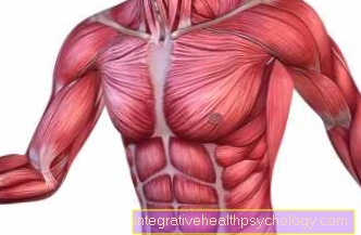
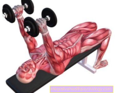
The pectoralis major muscle (large chest muscle) arises near the breastbone on the chest and attaches to the upper arm. Its functions are the adduction of the arm from the raised position, the internal rotation of the arm and the anteversion.
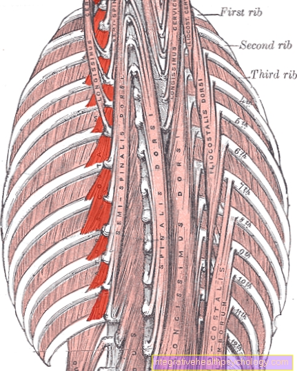
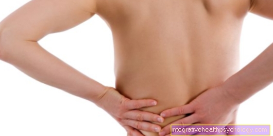
The iliac-rib muscle (Musculus iliocostalis) extends on the back from the iliac bone to the neck and belongs to the autochthonous back muscles. Its tasks are the straightening and stabilization of the spine with paired contraction, with one-sided contraction
The external jaw muscle (pterygoideus lateralis muscle) is the only jaw opener in the human jaw. It arises from the sphenoid bone and attaches to the lower jaw bone.
The adductor magnus is the largest muscle in the adductor group. It runs from the pubic bone to the lower thigh bone, where it forms the adductor canal and its main function is to bring the thigh towards the
The half-tendon muscle (M. semitendinosus) belongs to the rear thigh muscles and extends from the ischial tuberosity to the inside of the knee joint, where it attaches to the shin. He stretches in the hip joint and bends in the knee joint.
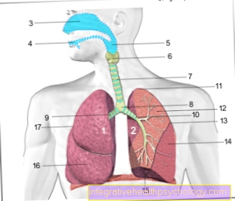
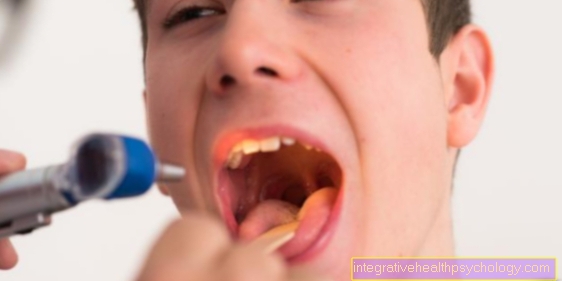
The throat connects the mouth and nose with the trachea or esophagus. The 15 cm long muscle tube is used to transport both breathing air and food through the swallowing reflex. The throat can be divided into three sections, with these through
The inner wing muscle (musculus pterygoideus medialis) belongs to the masseter muscles. It arises on the sphenoid bone, attaches to the lower jaw bone and causes the jaw to close. Besides, it also helps in the grinding of the food by holding the lower jaw
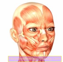
The back of the head and forehead muscle (musculus occipitofrontalis) belongs to the facial muscles and pulls the eyebrows upwards. This creates wrinkles on the forehead, also known as frowns.
The semi-membranous muscle (M. Semimembranosus) belongs to the ischiocrural muscles and lies on the back of the thigh. Its functions include flexion in the knee joint and extension in the hip joint.
The testicle lifter (M. Cremaster) consists of muscle fibers from the abdominal muscles. The muscle fibers follow the spermatic cord and attach to the testicular cover. The name corresponds to the function: the testicle lifter pulls the testicle closer to the abdominal wall like a protective reflex
The external oblique abdominal muscle (Musculus obliquus externus abdominis) is the largest and most superficial abdominal muscle in humans. Its function consists in tilting the axial skeleton, which can be trained very well with lateral push-ups.
The comb muscle (M. pectineus) connects the pubic bone with the thigh bone and belongs to the adductor group of the thigh. Its functions include flexion, external rotation and adduction of the thigh. In athletes it is often of
The upper eyelid lifter (M. levator palpebrae superiores) is an external eye muscle that is counted among the mimic muscles. When it contracts, the eye opens. If the muscle is damaged, what is known as ptosis can occur
The short adductor muscle (M. adductor brevis) belongs to the adductor group of the thigh and brings the thigh closer to the body. He can often be affected by the torn muscle fiber.
The rib muscles (Mm. Levatores costarum) belong to the trunk muscles. Its origin is the transverse process of the last cervical vertebra and the 1st to 11th thoracic vertebrae and their attachment to the underlying ribs. Their function is to lift the ribs, thereby creating
The temporal muscle (Musculus temporalis) belongs to the masticatory muscles of the human jaw and is the strongest jaw closer. It arises from the temporal fossa and attaches to the lower jaw.
The bronchial tree fills the lungs, emanates from the trachea, and undergoes 22 divisions before it joins the alveoli. The bronchi, the air-conducting paths, get smaller and smaller from top to bottom. Especially in winter it often happens in the frame
The masseter muscle originates from the anterior zygomatic arch, attaches to the mandibular angle and, together with the temporalis and medial pterygoid muscles, closes the jaw. This creates pressure on the salivary drainage
The temporoparietal muscle (temporoparietalis muscle) belongs to the facial muscles, runs on both sides of the skull and spans the galea aponeurotica, a tendon plate that serves as an attachment to several facial muscles.


.jpg)
.jpg)

.jpg)
.jpg)

.jpg)